An 8-year-old spayed female Miniature Schnauzer dog was initially presented for evaluation of polydipsia. A blood chemistry initially showed only mildly elevated ALP activity. She was treated for Leptospirosis with doxycycline. On re-evaluation, abdominal pain and left renomegaly were present. Urinalysis revealed low specific gravity (1.022), proteinuria, hematuria, and bacteruria. Abnormalities on serum biochemistry included azotemia and elevated ALP activity.
An 8-year-old spayed female Miniature Schnauzer dog was initially presented for evaluation of polydipsia. A blood chemistry initially showed only mildly elevated ALP activity. She was treated for Leptospirosis with doxycycline. On re-evaluation, abdominal pain and left renomegaly were present. Urinalysis revealed low specific gravity (1.022), proteinuria, hematuria, and bacteruria. Abnormalities on serum biochemistry included azotemia and elevated ALP activity.
Renomegaly: neoplasia, abscess, pyelonephritis, hydronephrosis, ureterolith, renolith, or granuloma.
The left kidney contains a proximal ureteral calculus with severe pyoureter and pyelonephritis pattern. It is also possible that the left kidney may be neoplastic given the nodular change in the renal pelvis.
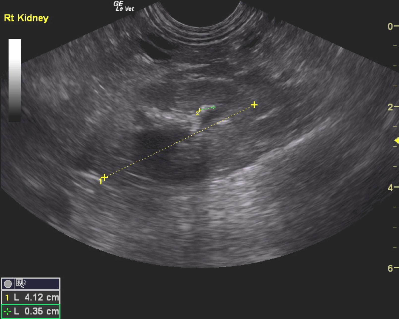
Renal calculi are noted in the right kidney as well; however, only minor degenerative changes were noted without evidence of obstructive disease.
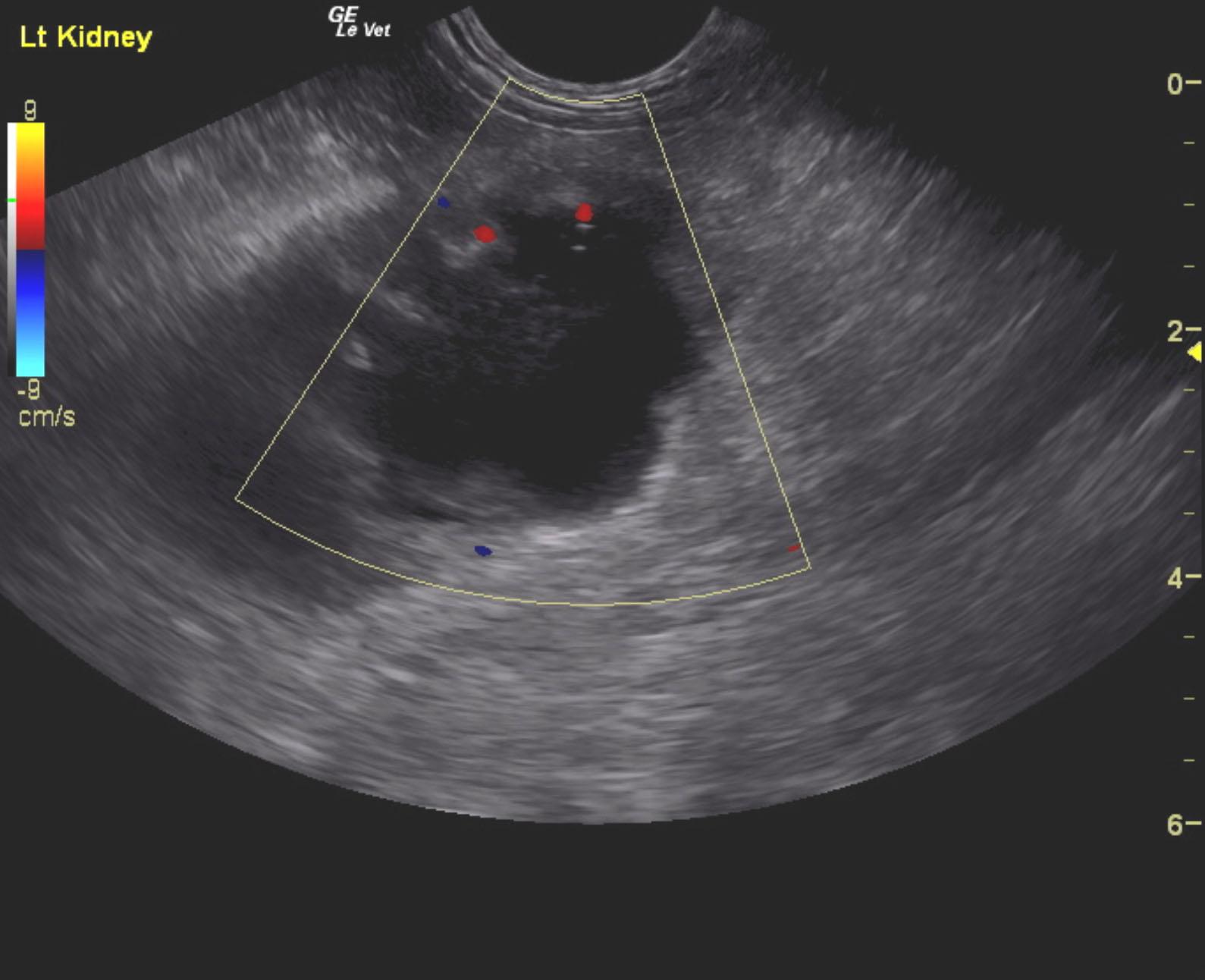
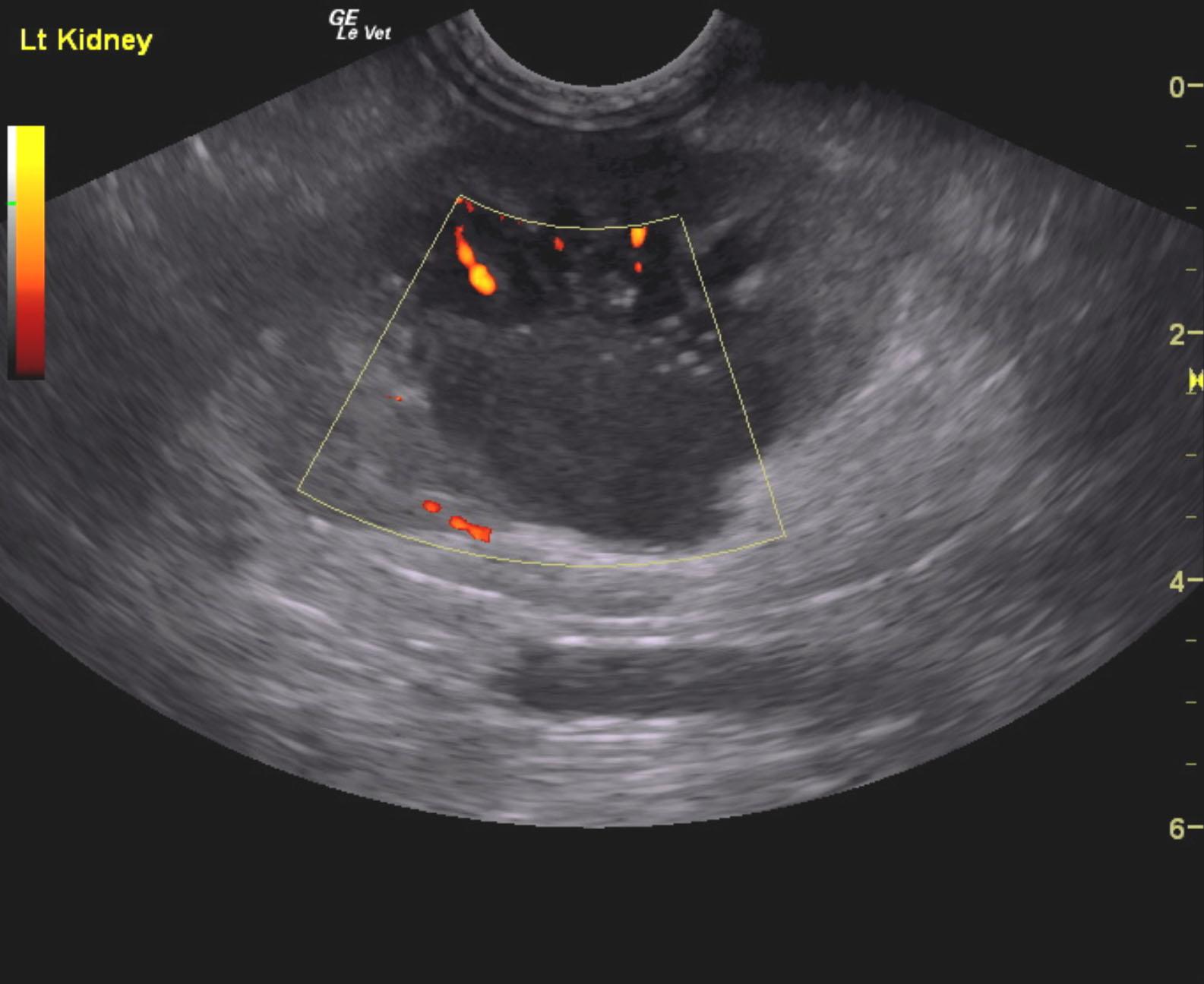
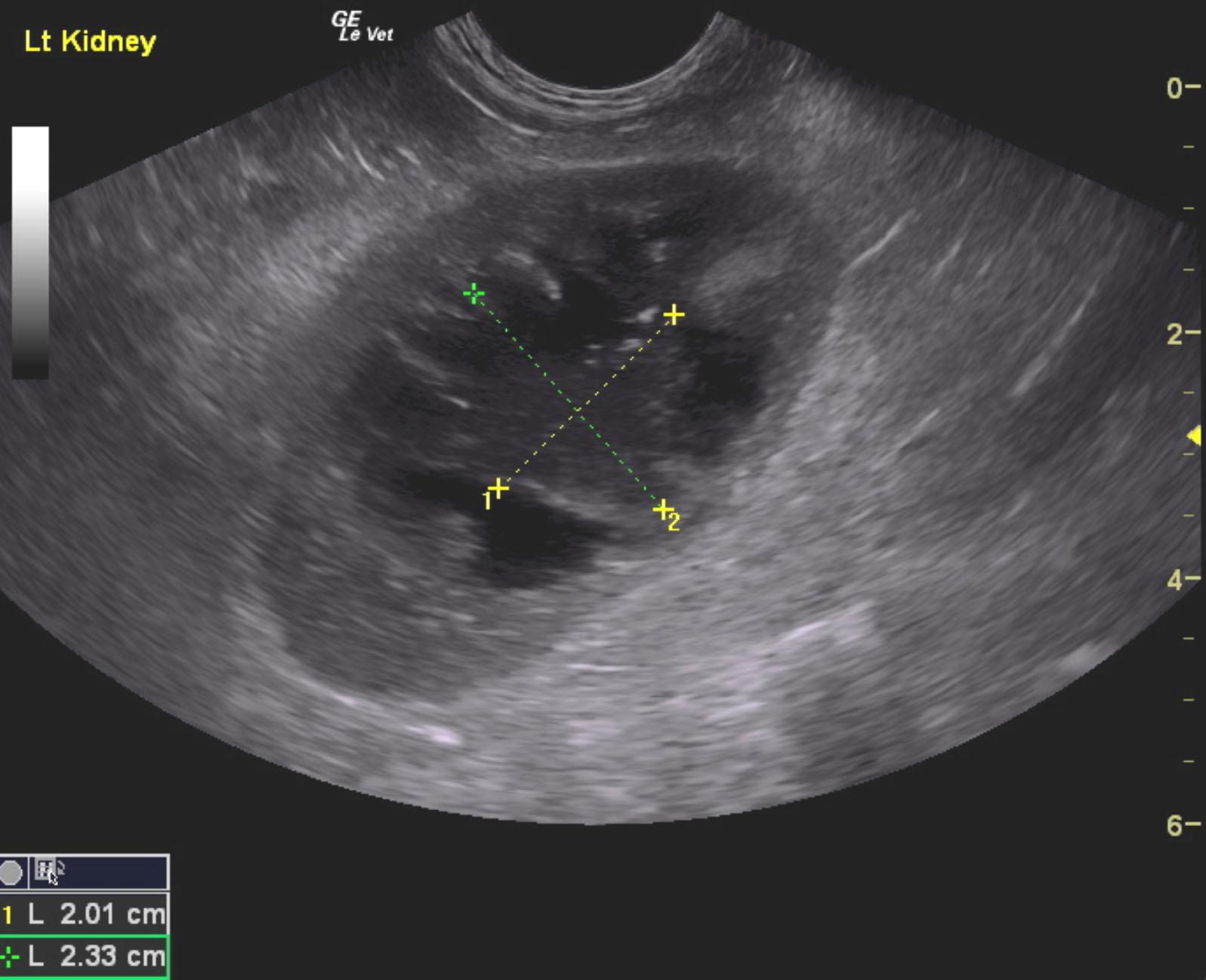
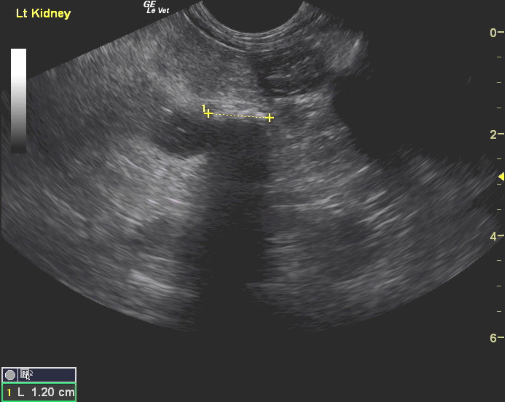
The left kidney in this patient presented severe hydronephrosis with echogenic debris, which is most consistent with severe pyelonephritis. Pericapsular inflammatory pattern was noted, along with embedded calculus at the corticomedullary junction. A calculus was embedded at the proximal ureter of the left kidney, and measured 1.2 cm. The left ureter was dilated and filled with echogenic debris measuring 1.1 cm. The left renal pelvis measured 1 x 3 cm with a nodular change disrupting the renal pelvis. This nodule measured 3.03 x 2.2 cm, and could represent an abscess or tissue proliferation. The right kidney presented mild degenerative changes with no specific pathology. The right kidney measured 4.12 cm with non obstructive corticomedullary calculi. A slight amount of free fluid was noted in the caudal abdomen. This is likely due to inflammation associated with left ureteral and left renal pathology.