An 8-year-old SF DSH was presented for evaluation of anorexia for 3 days and lethargy for approximately 1 week. Abnormalities on CBC and serum biochemistry were anemia, leukocytosis, and azotemia. On survey radiographs, hydronephrosis with ureteroliths was evident.
An 8-year-old SF DSH was presented for evaluation of anorexia for 3 days and lethargy for approximately 1 week. Abnormalities on CBC and serum biochemistry were anemia, leukocytosis, and azotemia. On survey radiographs, hydronephrosis with ureteroliths was evident.
Ureterolith with obstructive uropathy
Renal – chronic kidney disease, neoplasia, renoliths, pyelonephritis
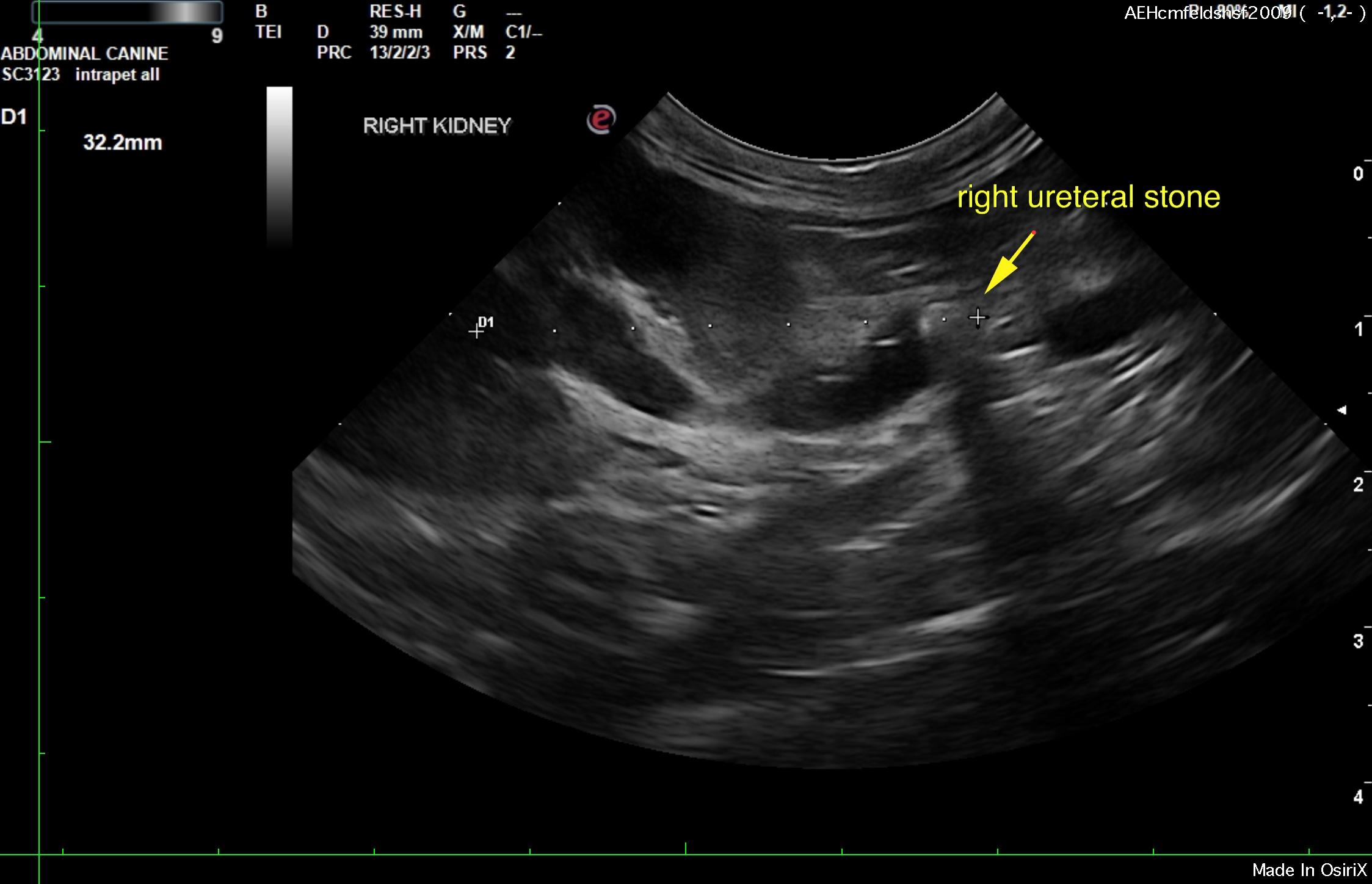
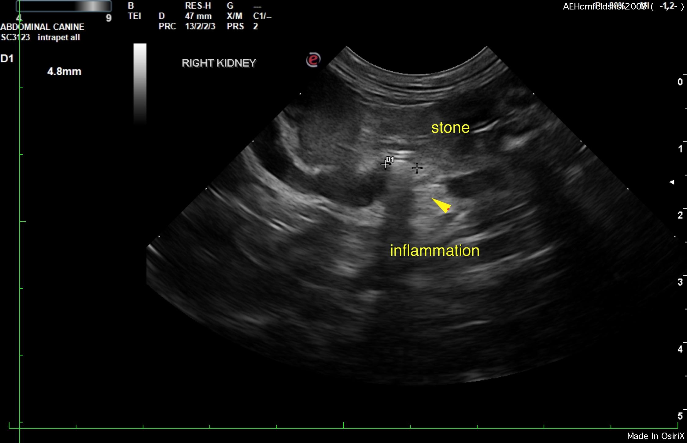
Right hydroureter was noted and measured 0.42 cm in dilation. The right kidney measured 4.01 cm with corticomedullary mineralization and loss of corticomedullary definition. Slight pyelectasia was noted. Calculus obstruction was noted 3.2 cm caudal to the right renal pelvis. The calculus measured 0.4 cm. An inflammatory pattern was noted around the ureteral obstruction along with embedded calculus. The right renal pelvis presented debris suggestive for concurrent pyelonephritis. The left kidney presented mild degenerative changes, cortical infarcts and collapse measuring 3.7 cm with moderate degenerative changes. Both kidneys presented solid color flow signals in the renal cortices.
I recommend referral for surgical or interventional radiology procedure for removal of the right ureteral stone. Urine culture and sensitivity as well as IV fluid protocol to treat for acute renal failure would be recommended in the meantime. The kidneys do appear to be significantly viable, yet the obstruction and any infection as well as complicating issues such as hypertension should be addressed.