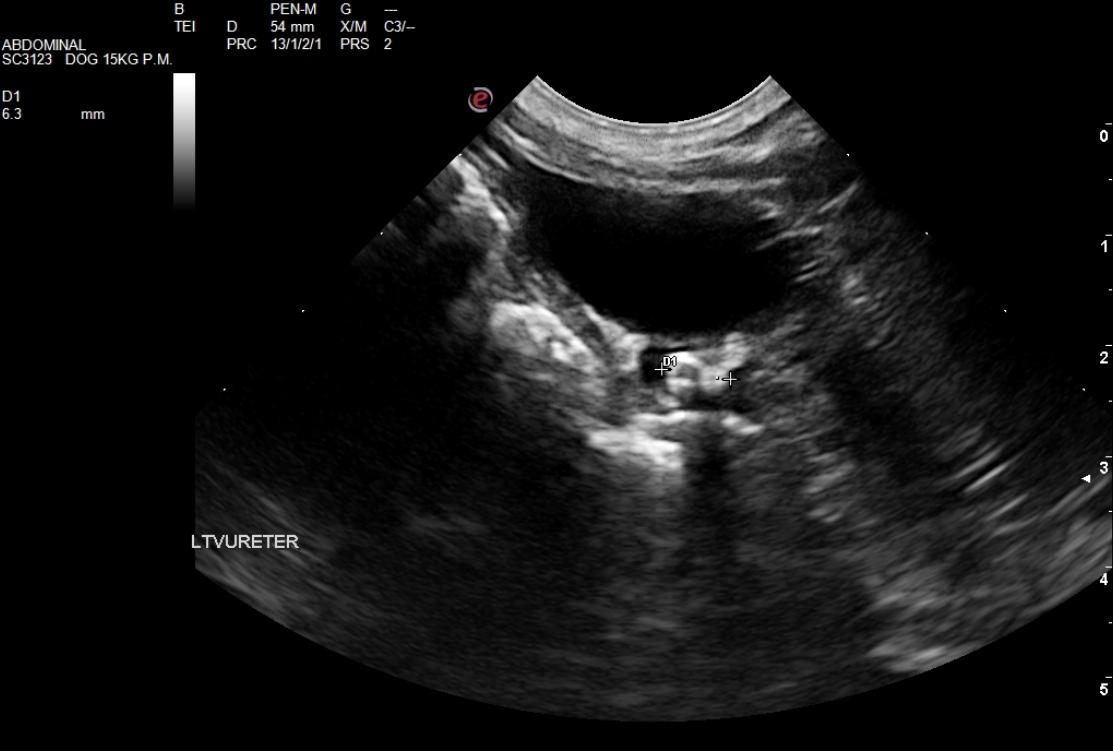
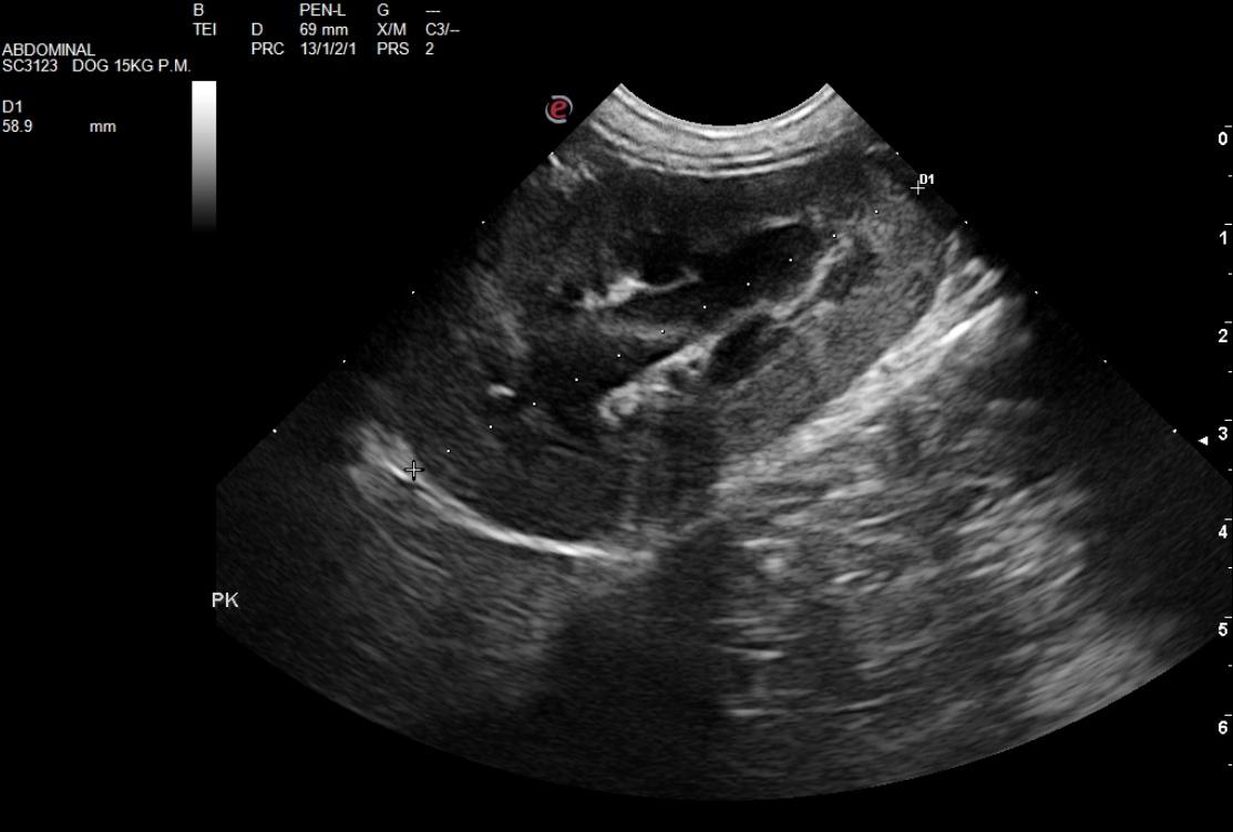
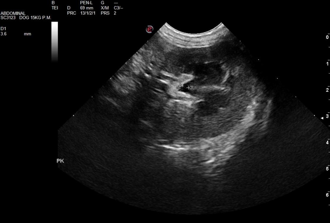
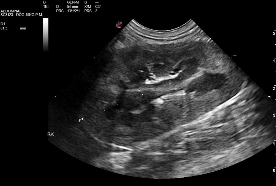
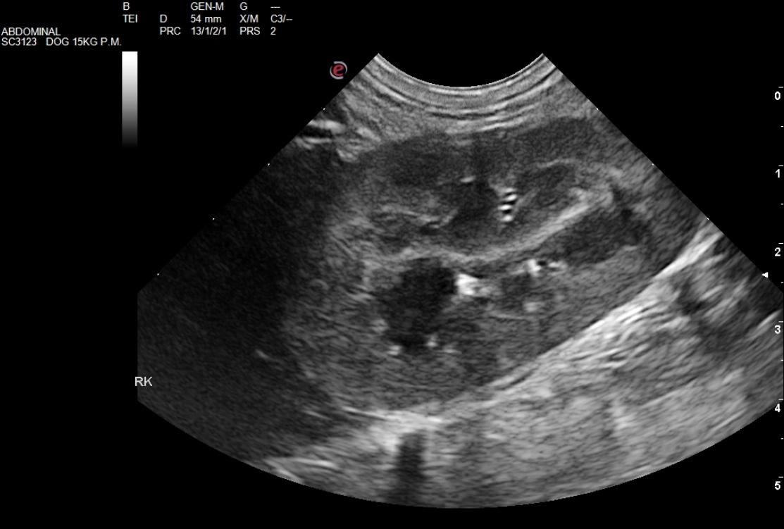
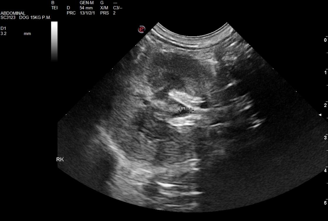
The patient is a spayed female canine Rottweiler mix, 19 weeks old who had a previous visit to emergency veterinarian for urinary problems. On physical the patient had slight dehydration but was otherwise normal. UA found no infection. Patient sent home on Rimadyl. CBC: two days ago: Anemia of 28.8, normocytic, normochromic. Azotemia (BUN = 34, CRE = 1.6) Hyponatremia (Na = 135) today: Anemia of 35. Improvement in BUN and CRE values (27 and 1.3 respectively), Hypoalbuminemia. Sodium back up to 145. UA: SG = 1.015, Glycosuria, proteinuria, pH=6
The patient is a spayed female canine Rottweiler mix, 19 weeks old who had a previous visit to emergency veterinarian for urinary problems. On physical the patient had slight dehydration but was otherwise normal. UA found no infection. Patient sent home on Rimadyl. CBC: two days ago: Anemia of 28.8, normocytic, normochromic. Azotemia (BUN = 34, CRE = 1.6) Hyponatremia (Na = 135) today: Anemia of 35. Improvement in BUN and CRE values (27 and 1.3 respectively), Hypoalbuminemia. Sodium back up to 145. UA: SG = 1.015, Glycosuria, proteinuria, pH=6