-
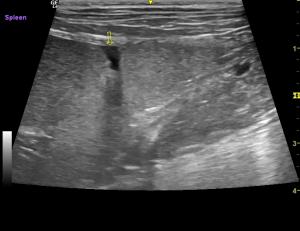
Small tear in the spleen?

11 yo MN Labradoodle 57# U/s for asymptomatic mild elevated ALP before dental. Normal abdomen except for a 5mm apparent tear in the spleen, with scant amount of free fluid and omentum patching the area. How often does this happen? The dog is doing great, normal HCT, no hx of trauma. I rechecked the spleen…
-
PDA?
8 week old shih-tzu with loud left-sided systolic murmur, no clinical signs reported although I believe there was some pulmonary edema by the end of my exam. -In the right – sided short axis view of the MPA, I do see turbelence under the bifurcation of the PA but the entire PA is not turbulent. …
-
Suspected salivary mucocele?
13 yrs MN Bichon mix 5″ x 3″ soft fluctuant swelling near the R submandibular LN Would this be consistent with a salivary mucocele? No CF on the top structure, mild CF on the gland below. I was there for an echo and was curious what this potential mucocele would look like. Thank you! Karen
-
liver tru cut biopsy
Hi all, I have a patient, 8 y.old, MN, JRT with diffuse changes in a relatively small liver and suspected microvascular dysplasia (no PSS shunt seen). I am going to take a tru cut biopsy but I am wondering, with these diffuse changes, if I should take just a single biopsy or more? I imagine…
-
Caecum?
7 year old medium sized cross breed with vomiting There was an area of shadowing within intestine adjacent to ileocolic junction, is this caecum? There was a surrounding lymphadenopathy and a tiny amount of free fluid.
-
Unusual mass
12 year old Lab with inappetance, weight loss, anaemia and ascites Large (10cm+) mass in right cranial quadrant Can you tell where this mass is coming from please?
-
sternal mass – biopsy suggestions
i’m planning to biopsy this mass with ultrasound guidance tomorrow. Was planning to sedate and use an 18 GA needle. Would you do anything differently?
-
Kidneys with micro infarcts (?) and liver mass in a hypertensive, hyperthyroid cat
15 year old MN DSH with blood pressure >200, mildly elevated T4 and Ca with weight loss. Still eating and no vomiting or diarrhea. Could you please give your impression of the kidney and liver?
-
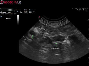
Adrenal gland images

First 2 images and videos are now, 3 months post adrenalectomy The third video and following image is 3 months ago when right adrenal mass first diagnosed
-
Small intestinal inflammation
Can you explain what is happening here with the mucosa in a dog with diarrhoea? Luminal section is hyperechoic and luminal interface is blurred with distinct change to outer hypoechoic mucosa.
