 This is one of those issues which is probably going to make me look a complete beginner but….
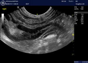
Images (all dorsal plane from R side) from an 18 m.o. MN Visla with a history of daily (early mornings most days) vomiting for 2 months. No other signs, no weight loss, otherwise 100% well in self, biochem/haem unremarkable. Abdominal ultrasonography otherwise unremarkable apart from the fact that the duodenum has widespread patchy/linear hyperechoic changes of the type often seen with dilated lacteals. And there is a slightly large duodenal lymph node.
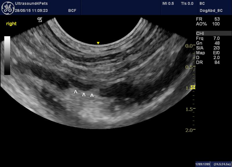
There is a striking, discrete discontinuity in the anti-mesenteric mucosa of the descending duodenum where the hyperechoic luminal content appears to ‘track’ into the mucosa.
However…there’s no real reaction outside the serosa, the surface of the defect is not hyperechoic and the adjacent duodenal wall is not thickened as I would expect adjoining an ulcer. It’s on the wrong aspect of the duodenum for the normal position of the duodenal papillae and associated ducts. I am a bit wary of over-interpreting irregularities in the wall of the proximal duodenum especially around the cranial flexure where I often see some folding of the mucosa in normal dogs.
So, I’d really welcome some input on these images -pathological or normal?

16 responses to “duodenal ‘changes’ in a vomiting 18 m.o. Viszla dog”
….could be a Peyer’s
….could be a Peyer’s patch???
….could be a Peyer’s
….could be a Peyer’s patch???
Nice eye UFP, these are
Nice eye UFP, these are mucosal striations which are important because sometimes you see them before a PLE starts so watch the albumin. We usually see them with PLE cases. There’s a fine line between mucosal speckling and striations but these look like striations. So Lymphangectasia is on the front concern here. Givent he age look for parasites and blanket tx fenbendazole metro. Could start treating ibd/PLE with diet purina HA or royal canine HP. Peyers patches will be in the ileum but good thought.
Technically could scope and do a corn oil test to dilate the lacteals prior to scope and duodenal bx.
Check out the basic search on mucosal striations:
http://sonopath.com/members/case-studies/search?text=mucosal+striations&species=All
Thanks as always Eric,
Right,
Thanks as always Eric,
Right, I’ve added another image of the same piece of descending duodenum. It’s not the striations that are at issue -it’s the ‘crater’ (arrowheads) and the hyperechoic ‘tracts’ leading off deeper into the wall that I’m wondering about.
I’ve seen some debate about irregularities in the duodenum:
http://www.sid.ir/en/VEWSSID/J_pdf/11592008S07.pdf
(page 58, first paragraph -it’s OK it starts at page 56!)
Yes, Peyer’s patches mostly ileal but sometimes duodenal…..
http://www.ncbi.nlm.nih.gov/pubmed/1595185
what’s an eye UFP?
Best regards
Roger
Short for Ultrasound4Pets
Short for Ultrasound4Pets 🙂
I see the striations- but all I see is ingesta.
I guess a cine is always good to see what remains after the ingesta moves.
I don’t get a clear view of the ‘anti-mesenteric” wall.
Thanks as always Eric,
Right,
Thanks as always Eric,
Right, I’ve added another image of the same piece of descending duodenum. It’s not the striations that are at issue -it’s the ‘crater’ (arrowheads) and the hyperechoic ‘tracts’ leading off deeper into the wall that I’m wondering about.
I’ve seen some debate about irregularities in the duodenum:
http://www.sid.ir/en/VEWSSID/J_pdf/11592008S07.pdf
(page 58, first paragraph -it’s OK it starts at page 56!)
Yes, Peyer’s patches mostly ileal but sometimes duodenal…..
http://www.ncbi.nlm.nih.gov/pubmed/1595185
what’s an eye UFP?
Best regards
Roger
Short for Ultrasound4Pets
Short for Ultrasound4Pets 🙂
I see the striations- but all I see is ingesta.
I guess a cine is always good to see what remains after the ingesta moves.
I don’t get a clear view of the ‘anti-mesenteric” wall.
That looks like the accessory
That looks like the accessory pancreatic duct to me… curvilinear pattern maintained.
That looks like the accessory
That looks like the accessory pancreatic duct to me… curvilinear pattern maintained.
good, that makes sense.
I
good, that makes sense.
I just found another reference that says that, sometimes, the two lobar pancreatic ducts can enter the duodenum separately -maybe that’s what we’re seeing (there appears to be two, one running cranially from the ‘crater’ and one going more caudally).
I thought it was a bit unusual for it to be on the lateral (abaxial, antimesenteric) aspect of the duodenum but maybe just a bit of individual variation or maybe not a completely dorsal plane view.
good, that makes sense.
I
good, that makes sense.
I just found another reference that says that, sometimes, the two lobar pancreatic ducts can enter the duodenum separately -maybe that’s what we’re seeing (there appears to be two, one running cranially from the ‘crater’ and one going more caudally).
I thought it was a bit unusual for it to be on the lateral (abaxial, antimesenteric) aspect of the duodenum but maybe just a bit of individual variation or maybe not a completely dorsal plane view.
great info… yes I have seen
great info… yes I have seen them on this side as well on occasion. Bottom line not pathological and yes a clip would define it better as well.
great info… yes I have seen
great info… yes I have seen them on this side as well on occasion. Bottom line not pathological and yes a clip would define it better as well.
Thank you both.
I see what
Thank you both.
I see what you are referring to now. If I just looked at the 3rd image I would have thought that to be ingesta. Each day is a new learning experience for me.
Just a note about the history. I have seen dogs that I consider to have “gastro-esophageal” reflux. Those dogs seems to always vomit in the morning. I have seen some response with an H2 blocker like a Pepcid AC (10 mg probably OK on this dog) before bed and feeding a small meal before bed.
Many of those dogs will stop vomiting.
Just a thought
Thank you both.
I see what
Thank you both.
I see what you are referring to now. If I just looked at the 3rd image I would have thought that to be ingesta. Each day is a new learning experience for me.
Just a note about the history. I have seen dogs that I consider to have “gastro-esophageal” reflux. Those dogs seems to always vomit in the morning. I have seen some response with an H2 blocker like a Pepcid AC (10 mg probably OK on this dog) before bed and feeding a small meal before bed.
Many of those dogs will stop vomiting.
Just a thought