
 Pancho 11 yr, MN, DSH obese patient being evaluated for thrombocytopenia found on pre op bloodwork for dentistry.
Bloodwork and virals normal except for an abnormal fPL
Echo showed VPC’s on the ECG but no significant changes on the heart structure.
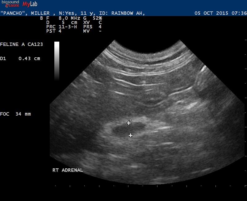
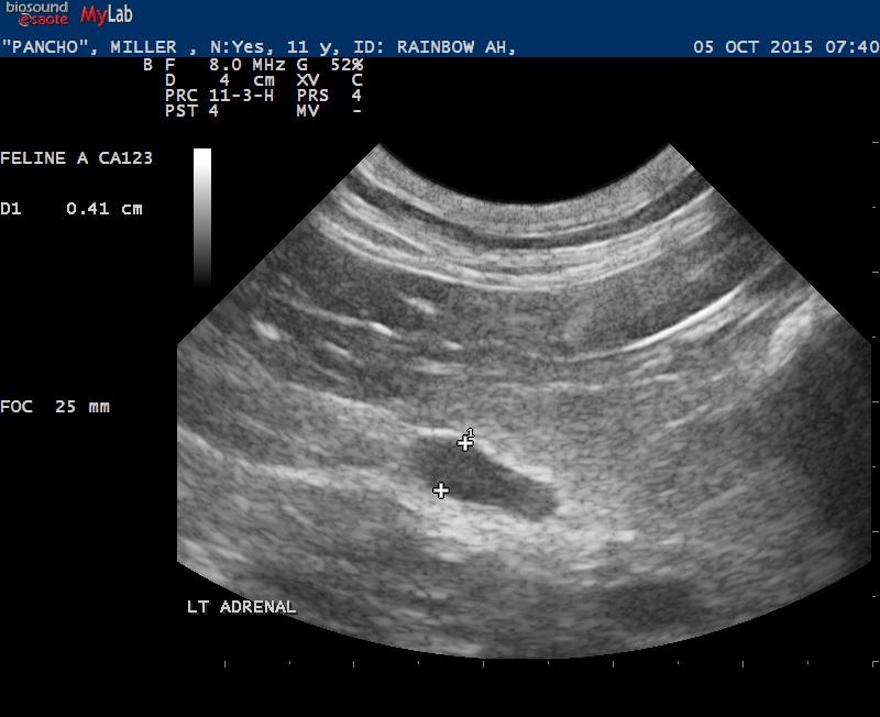
Am curious as to what you think of the adrenal glands. On the abdomen exam it seemed there was inflammation around the adrenals with the hyperechoic fat.
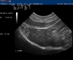
Also the liver lesion – biliary cystadenoma?
Thanks for the input.
Okay added the other images, forgot to hit the insert button. I didn’t have a cineloop on the liver lesion but my impression was it was in the liver.
8 responses to “Feline thrombocytopenia”
Have any cine’s? Could the
Have any cine’s? Could the nodule be coming off the diaphragm?
Only image loaded is a still
Only image loaded is a still image of the liver with a hyperechoic nodule in the region of the diaphragm so cannot comment on the adrenals. Liver mass could be neoplasia, granuloma, resolved abscess, organized hematoma. Does the cat show clinical signs of thrombocytopenia and has it it been confirmed – repeat CBC, peripheral blood smear evaluation?
Yeah the CBC has been
Yeah the CBC has been repeated and the path review indicates significantly decreased platelets. Not clincial other than mild brusing with previous cystocentesis.
Amy, I had a similar looking
Amy, I had a similar looking hepatic nodule located next to the GB in a cat I scanned last week. I had recommended FNA but did not get to do it.
Yeah that is pretty identical
Yeah that is pretty identical 🙂
Older cats get cystadenomas
Older cats get cystadenomas that look like this.. They can transform into adenoca but when they are passive…i.e. just look like blotches on a wall as opposed to holes in the wall then im less concerned… FNA usually unrewarding as these are fibrous structures and need core bx for the dx. If early hepcell carcinoma then fna can give the dx. But in animage’s image the nodule is adjacent to the diaphragm but doesnt move it at all. The D stays in its contour. This is good. Bad things next to structures cause mini mass effects and deviate the D or in Pankatz case would deviate the Gb or cystic duct and that isnt happening either. Good to watch these lesions in 30 days making sure they stay put and dont grow or sample but these look like typical older cat lesions to me…. cystadenomas and occasional lipogranulomas but if they start doing the mass effects that I speak of or grow then I pay more attention to them.
What do you think of the
What do you think of the adrenal glands? I did get those images uploaded.
Thank you!
These adrenals are feline
These adrenals are feline stressed glands… try scannign a CRF cat or hyperthyroid cat they will look like this,,, rounded, hypoechoic, hypersecretory and jump on the probe.