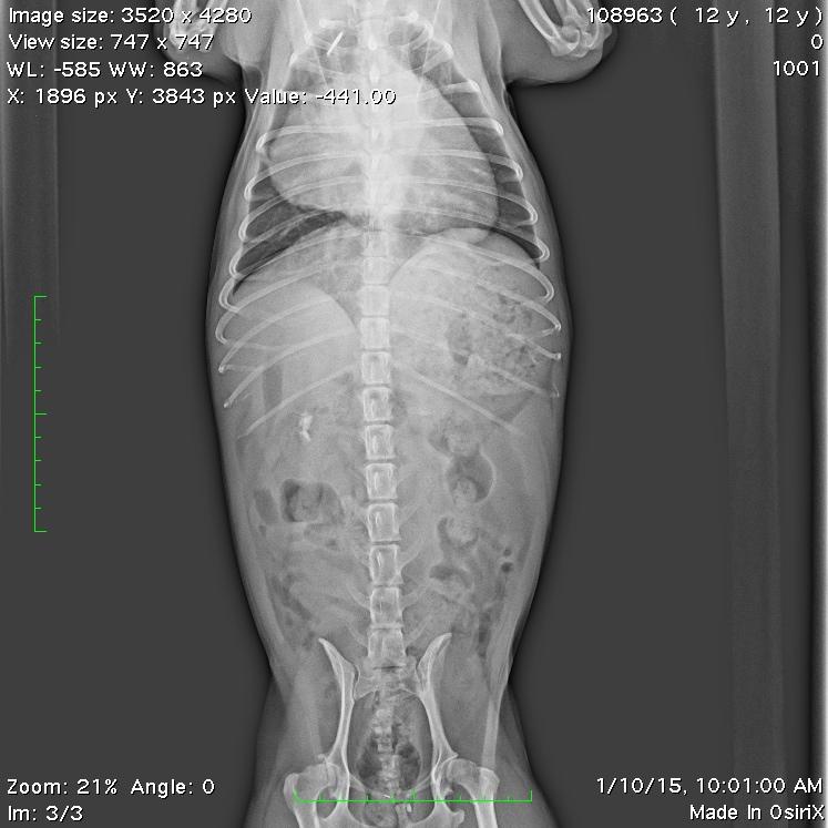
Honey is a 12 1/2 year old Pomeranian that had a TCC of the L kidney. The kidney was removed about 1 year ago and the histopath indicated the mass was incompletely removed.
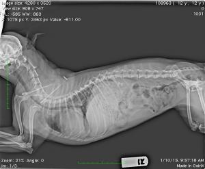
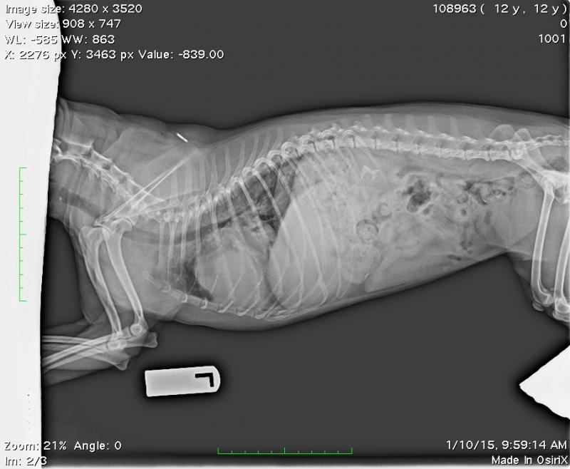
Honey has a cough that has been previously diagnosed as a collapsing trachea. I have verified with x-rays that she does indeed have a collapsing trachea. The cough recently has gotten worse and the quality of the cough has changed. The x-rays revealed a mass involving the R medial lung lobe.
Honey is a 12 1/2 year old Pomeranian that had a TCC of the L kidney. The kidney was removed about 1 year ago and the histopath indicated the mass was incompletely removed.
Honey has a cough that has been previously diagnosed as a collapsing trachea. I have verified with x-rays that she does indeed have a collapsing trachea. The cough recently has gotten worse and the quality of the cough has changed. The x-rays revealed a mass involving the R medial lung lobe.
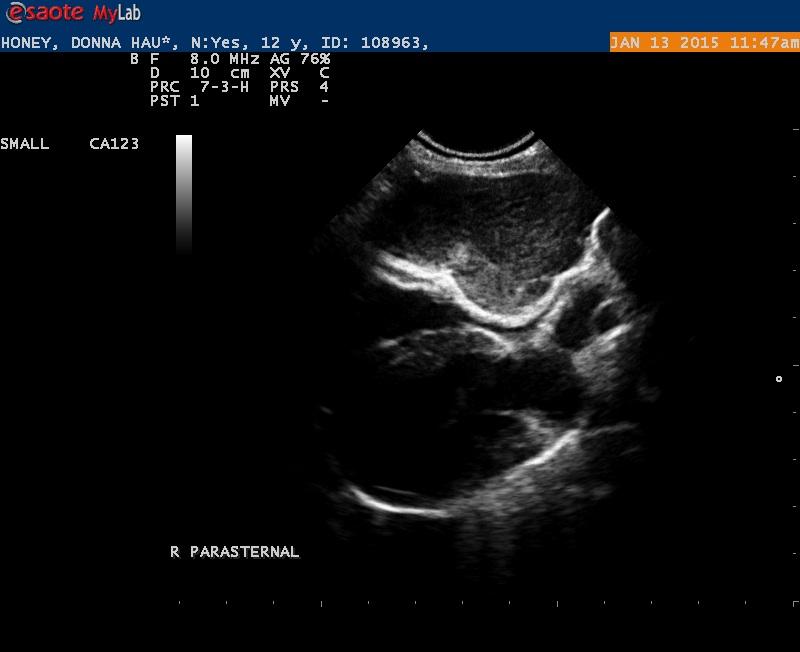
I did an echo today and I did indeed confirm a large mass. The ? – how do you know if this is of pulmonary orgin compressing the R side of the heart- or is it a mass associated with the heart.
On the R parasternal images it appears to be outside the pericardium and compressing the right side of the heart.
L sided function remains fine. No murmurs were heard.
Could this mass be a TCC?
I wanted to stick a needle in the mass- but the owners declined. They don’t want to put Honey through any more treatment.
Clinically Honey is doing great. I will post the x-rays, 1 .jpg and 1 cine – all from the right parasternal. I saw the mass best from this view.
4 responses to “Heart Base Mass”
I doubt this is TCC as it
I doubt this is TCC as it would be the oddest met pattern for TCC that I have seen but of course without a needle can’t really know.
I believe this mass to be outside the heart or deriving from the pericardium or attached to the pericardium for this reason:
Count the lines… my arrows smallest to largest show the RV, epicardium, pericardial space (slight fluid), and pericardium… then the mass. Boy I’m dyin to put a needle in that too randy:) Connective tissue tumor or something slow growing suspected. Wrong position for ao body tumor/chemodectoma but its the right breed.
Thanks EL
Thanks EL
Thanks EL
Thanks EL