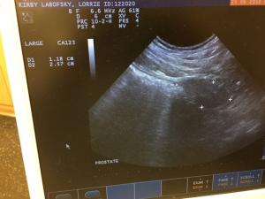
Kirby is a 10 year old Lab with a history of hematuria. Per owner no history of dysuria/poilakuria.
Last Urinalysis had no blood and the urine culture was negative.
I did an ultrasound on Kirby and could find no lesions in the bladder or prostate.
I did think there was some shadowing in the L kidney- maybe a stone producer?
No history of pain that might account for stone movement.
I am posting 2 cines of the kidneys and a .jpg of the prostate. No images of the bladder- looked totaly normal.
Any feedback would be appreciated.


8 responses to “Hematuria”
Renal stone is most likley
Renal stone is most likley incidental. With the hematuria and normal bladder/prostate and no hematuria on urinalysis, would think of lower urethra/prepuce etiology. May require contrast urethrogram and if available urethroscopy.
Thanks Remo
Thanks Remo
You have eveidence of mild
You have eveidence of mild pyelectasia on the second video. Now you need to look at why. I think that you need to examine the renal crest and it’s associated pelvis in greater detail. This is easiest in a transverse plane.Look for thickening of the wall of the proximal ureter . Also look for signs of double layering. Look to see how echogenic the fluid is in the renal pelvis if you can image it. You will need to zoom in to look at it better. Examine the crest to see if it is hyperechoic either in general or in focal diffuse patches. You may also want to measure an RI value. These are some observations that might help you identify an underlying pyelonephritis.
Thanks bhylands77.
Can you
Thanks bhylands77.
Can you post some images of the changes you describe. You sort of lost me at pyelectasia 🙂
Here is an example of the
Here is an example of the renal crest showing a flare from a case of pyelonephritis. The second view shows how regional it can be.
The other image is that of a
The other image is that of a zoomed view of the renal crest and proximal ureter. This is where you look for distinct double layering as seen with direct inflammation associated with an ascending infection.
Thanks for the images.
Thanks for the images. Clarifies what you were saying.
NICE BOB THANK YOU… I ALSO
NICE BOB THANK YOU… I ALSO GUESTIMATE INFECTION WHEN THE FLUID IS ECHOGENIC AND THE RENAL PELVIC FAT IS ILL-DEFINED. sorry for the caps:)
Pyelectasia can come from scarring as well as its common in stone movers so its important to take bobs comments along with everything else clinically becasue you can push abs for 6 weeks to tx pyelonephritis when the pyelectasia may be left over from scarring or fullness from moving stones historically and no infection… ive seen this with pyelocentesis of clean urine in pyelectasia cases. Aside from US guided pyelocentesis with a big enough dilation its educated guesswork often in these cases without a pelvic sample.