- 8 year old llasa mix (MC)
- 2 mo ago, episode of back pain, put on tapering dose of metacam x 9 days
- Now, acute onset vomiting bile, icterus
- No other previous dz
- Blood work (normal 2 mo ago): ALT>2000, bili 7.0, ALP 250ish, BUN 9, alb normal, glucose 70. Urinalysis showed bili crystals but no other types of crystals.
- Rads: microhepatica, stenosis at T13-L1
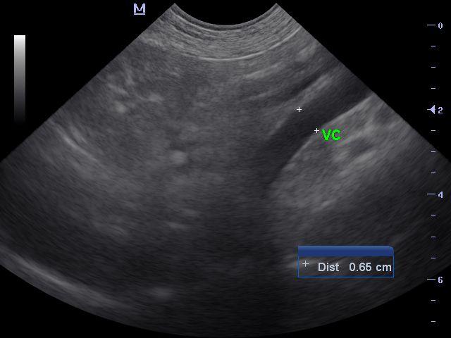
Scan: no urinary or renal mineralization, liver very small esp on left lobes. Gall bladder NSF, panc NSF. See attached video, unfortunately doppler disappeared into ether but I will try to explain.
The PV:VC had an approx 1:1 ratio (0.61 cm/0.65 cm), although VC diameter changed with pulse so this was measured at most dilated point. The aorta measured approx 0.95 cm.
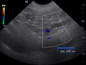
The PV seemed to originate in directly next to the VC then turned ventrally and appeared slightly dilated (and blue on doppler) then coursed caudally and exhibited mild mosaic pattern.
I am not sure if this just a portal vein taking a tortuous route, or if this could be a shunt. Sorry there’s no good doppler image, but the entire vessel was blue, and the part where the vessel moved dorsally did not have any mosaic.
Your thoughts appreciated.
Liz
16 responses to “High ALT, bili, and small liver”
I am not certain about the
I am not certain about the portal vein/vena cava for a shunt, but I can’t believe these symptoms are due to a shunt.
I would be more concerned about some sort of hepatopathy/hepatitis. I guess I would also be concerned about a post hepatic obstruction like a tumor or gall stone obstructing the CBD.
I am waiting to hear from others.
Tough to tell on these
Tough to tell on these views but that last one looks suspicious but given the chronic liver changes and small primary size portal hypoplasia and chronic hepatitis or acute on chronic more likely. Stones and swoillen kidneys should have been present with a primary shunt for 8 years with rare exception. For more shunt views check out the sonopod casts (http://sonopath.com/resources/instructional-library and tsearch shunt on the general search (http://sonopath.com/members/case-studies/search).
I would treat for acute phase disease including lepto and treat for liver failure. Needs liver bx for sure. The shunt part isnt important in this case til you get him out of failure then can submit more views of the portal branching with doppler. Along with iv ampicillin covering for lepto i would try this protocol:
Hepatic Support For Bile Acid Elevation +/- Hepatic Encephalopathy
Royal Canin Hepatic Support diet or Hills L/D, Metronidazole (7.5 mg/kg PO bid)over the next 14 days, Lactulose (Oral: 3.1-3.7 g/5 ml lactulose in a syrup base) long term to target 2-3 soft stools/day, with a high quality protein supplement of minor amount of yogurt or cheddar cheese. Monitor bile acids, with attention paid to dropping albumin, BUN or cholesterol. SAMe and neutraceuticals as needed. Urosodiol (10-15 mg/kg p.o.q24h) can be considered as hepatoprotectant and to enhance bile flow. Zinc serum level keep between 200—500 ug/dl. If deficient then tx zinc acetate 1-3 mg/kg/day. Gi protect if anorexic
Yes I definitely didn’t think
Yes I definitely didn’t think the symptoms due to shunt given the Bili of 7. However was just trying to figure out if this appearance of the PV was a normal variant. Thanks for your input!
Yes I definitely didn’t think
Yes I definitely didn’t think the symptoms due to shunt given the Bili of 7. However was just trying to figure out if this appearance of the PV was a normal variant. Thanks for your input!
This dog’s gall bladder
This dog’s gall bladder appeared totally normal. Pylorus was normal. No evidence of obstruction. Why would the Bili be so high?
This dog’s gall bladder
This dog’s gall bladder appeared totally normal. Pylorus was normal. No evidence of obstruction. Why would the Bili be so high?
The bili elevation will
The bili elevation will happen for 4 reasons:
hemolytic: rule out with anemia
parenchymal disease : this is case here. Can be from any insult lepto toxin chronic or acute failure
post hepatic obstrction: mucocele, cbd tumor, plug. stone
salmonella infection will do this as well
Most likley an acute
Most likley an acute hepatopathy – severeley elevated ALT that was normal 2 months ago. Low-normal glucose also is more likely with acute hepatopathy. Exposure to Metcam a possible cause in this case but consider other toxins, bacterial, viral, Leptospira.
Unlikley that you are dealing with PSS or portal vein dysplasia as no pre-existing signs and normal previous bloods.
The dog died acutely
The dog died acutely yesterday morning after developing petechia on abdomen and before the clinic had time to give FFP.
The dog died acutely
The dog died acutely yesterday morning after developing petechia on abdomen and before the clinic had time to give FFP.
More evidence of acute
More evidence of acute hepatopathy as most likley developed a hemorrhagic diathesis.
More evidence of acute
More evidence of acute hepatopathy as most likley developed a hemorrhagic diathesis.