- 4 year old MN Akita, travelled from Korea, then Austria
- Presented with severe pyrexia and skin lesions/cellulitis of distal limbs (biopsy severe acute necrotising pododermatitis)
- Mild inflammatory leukogram, mild hypoalbuminaemia and mild hyperglobulinaemia, leishmainia negative
- Skin lesions partial response to abs but break through lesions, Pyrexia/lethargy/lesions responded to steroids
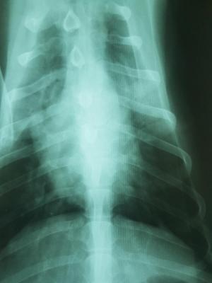
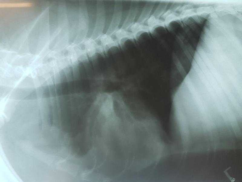
- Developed dyspnoea in hospital and coughing up frothy blood tinged fluid, initial rads (teaser image) and +5d rads following furosemide (other 2 images)
- 4 year old MN Akita, travelled from Korea, then Austria
- Presented with severe pyrexia and skin lesions/cellulitis of distal limbs (biopsy severe acute necrotising pododermatitis)
- Mild inflammatory leukogram, mild hypoalbuminaemia and mild hyperglobulinaemia, leishmainia negative
- Skin lesions partial response to abs but break through lesions, Pyrexia/lethargy/lesions responded to steroids
- Developed dyspnoea in hospital and coughing up frothy blood tinged fluid, initial rads (teaser image) and +5d rads following furosemide (other 2 images)
- I saw dog today for scan – heart normal, lung consolidated in peripheral nodules (aspirated) and cranial to the heart
- Could this be a lung lobe torsion? Any other input to this unusual case or scan images?
- Suspect skin lesions are immune-mediated secondary to some other pathology….
7 responses to “Lung consolidation”
The first videos look like
The first videos look like fat density around the heart but the last one is likely pneumonia. Maybe neoplasia but doubt it. 22 g fna for cyto and culture.
Thanks. Do you think I can
Thanks. Do you think I can exclude a torsion?
torsions in my experience
torsions in my experience have effusion associated with them and i dont see that here.
It’s the xrays that concern
It’s the xrays that concern me. And coughing up blood tinged effusion. The lung cranial to the heart also seemed consolidated but I know images are poor, I was missing my probe. I have sampled the peripheral nodules and advised CT so will keep you posted.
CT with contrast is the way
CT with contrast is the way to go here.
Results:
SITES:Consolidated
Results:
SITES:
Consolidated area of lung tissue, 4 direct smears
CYTOLOGICAL INTERPRETATION:
1. Moderate to severe mixed inflammation (pyogranulomatous)
2. Severe necrosis
3. Mild chronic haemorrhage
4. Mild fibroplasia
CYTOLOGICAL DESCRIPTION:
The cellularity was high in the cellular preservation adequate for review with
moderate background haemorrhage. There was a large amount of pale blue-pink,
moderately granular necrotic material dispersed throughout the smears. In some areas a
small amount of purple coloured fibrillar material was also evident (suspect mucus).
There was a large mixed population of inflammatory cells scattered throughout. The
most prominent cell types were medium to large, vacuolated macrophages and variably
preserved neutrophils. Few macrophages were showing erythrophagia. Other cell types
were fewer and consisted of occasional small to medium size lymphocytes. Occasional
plump spindle-shaped cells were seen singly throughout (interpreted to be reactive
fibroblasts).
COMMENT:
Anita, although microorganisms are not evident in these smears, infection is still
possible and aerobic bacterial and mycotic culture are recommended for further
characterisation. Other differentials for these findings include foreign body
inhalation and aspiration pneumonia. Neoplasia cannot be entirely ruled out; however,
this is considered less likely given the history supplied.
sounds like pneuomonia and
sounds like pneuomonia and necrosis to me. Any fungal in your region? should have seen fungal organisms wiht that sample though so thats down on the list. CT ideal here but I like baytril clindamycin on these, b-dilator and supportive care