– 4 month old FS DSH from a shelter with a history of on and off anorexia
– presented with fever and anorexia and tongue ulceration (suspect calici)
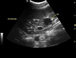
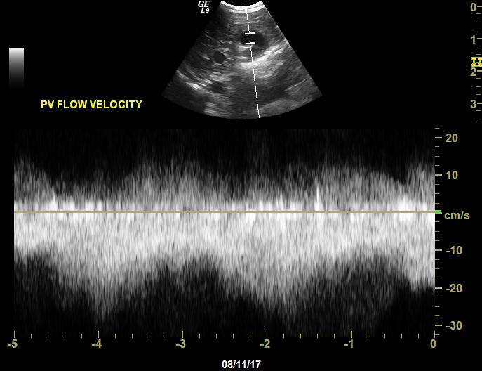
– abdominal u/s showed enlarged jejunal and medial iliacl LN’s’ – every thing else normal except for an enlarged and tortuous PV with high velocity flow
– I could not connect the PV to the CVC (or any other blood vessel ); the CVC appeared normal with laminar flow up until the diaphragm
– 4 month old FS DSH from a shelter with a history of on and off anorexia
– presented with fever and anorexia and tongue ulceration (suspect calici)
– abdominal u/s showed enlarged jejunal and medial iliacl LN’s’ – every thing else normal except for an enlarged and tortuous PV with high velocity flow
– I could not connect the PV to the CVC (or any other blood vessel ); the CVC appeared normal with laminar flow up until the diaphragm
– could this be some sort of shunt? PV anomoly? (in the last video, the CVC is incorrectly labelled PV fyi)
10 responses to “Odd PV in cat”
Im no expert for sure on
Im no expert for sure on shunts, but it appears in the last video that the PV is about the size of the CVC and Aorta until a short vessel empties into it with flow away from the probe. Where that is coming from I am not certain.
This looks like
This looks like agastriocaval shunt given the contour and position. From your video 3 position slide caudally about 1-2 cm keeping the focus on the cvc and look dorsally to it with the cf sector in that region. My bet is you see the shunt enter the cvc there at about 2 cm depth
Thank-you EL – will ty to get
Thank-you EL – will ty to get patient back on to take another look.
I am a bit confused here.
I am a bit confused here. Please clear this up for me.
It the portal vein dilated and torturous?
How does this happen when in most cases the shunt flows from the portal vein into the CVC or aorta.
Can you reference on Sonopath a drawing of this type of shunt.
Just trying to learn.
yes this is the diagram from
yes this is the diagram from the upcoming textbook (clinical approach to sonographic pathology) and the shunt hunt posters we are creating. Would need more views to confirm but the curve is that of a gastrocaval shunt or maybe gastroazygos. More images to see the post shunt pv would be needed as well but the pv will be small and tough to see after the shunt especially in a cat.
Thank you EL. That clearifys
Thank you EL. That clearifys the Gastic Caval Shunt. I understand.
Could you – if you have time identify the structures in a still I made of the final cine. Maybe I am interpreting the vessels incorrectly. This is a learning lesson for me.
Thanks
Randy
Randy, to clarify this
Randy, to clarify this picture, the dark blue vessel labelled PV was incorrectly labelled. It is in fact the CVC if that helps clarify things. This was a transverse view of the portahepatis. FYI I am rescnning this cat next week, so will see if I can get more images.
Jacquie
Randy, to clarify this
Randy, to clarify this picture, the dark blue vessel labelled PV was incorrectly labelled. It is in fact the CVC if that helps clarify things. This was a transverse view of the portahepatis. FYI I am rescnning this cat next week, so will see if I can get more images.
Jacquie
Totally. Please post any
Totally. Please post any pertinent images.
Thanks
Here you go Randy this should
Here you go Randy this should help:)