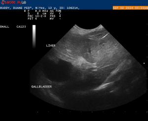
Buddy is a 12 year old Minature Poodle that was recently seen for weight loss, loss of appetitie and vomiting. Buddy has multiple medical issues including a protein losing enteropathy with an albumin of 1.9. Buddy is also a diabetic. I was doing a routine scan of Buddy’s abdomen and I believe there is a soft tissue mass in the gallbladder. I would like a second opinion. I am also including 1 image of the stomach. I would like some feedback on the gastric wall.
Buddy is a 12 year old Minature Poodle that was recently seen for weight loss, loss of appetitie and vomiting. Buddy has multiple medical issues including a protein losing enteropathy with an albumin of 1.9. Buddy is also a diabetic. I was doing a routine scan of Buddy’s abdomen and I believe there is a soft tissue mass in the gallbladder. I would like a second opinion. I am also including 1 image of the stomach. I would like some feedback on the gastric wall.


4 responses to “Polypoid Hyperplasia of the Gallbladder Neck”
I uploaded a number of
I uploaded a number of photos. I don’t know what happened to them. I will try again.
I uploaded a number of
I uploaded a number of photos. I don’t know what happened to them. I will try again.
Hi Randy this is not a
Hi Randy this is not a tissue mass but polypoid hyperplasia of th eGB neck. This is a common finding in older dogs and is typically benign but can cause secondary mucocele presentations but not in this case. Polypoid hyperplasia derives from the mucosa and is passive with respect to the CD, CBD, and GB wall so the GB CD and CBD maintain their contour as they are here (CBD not overtly seen but your views would have shown dilation had there been a cbd problem). Whereas a bliary carcinoma for example will cross through the wall of these structures and deviate contour and destroy mural architecture.
Check out the basic search on biliary carcinoma but this cat with a cbd carcinoma shows what I am describing
http://sonopath.com/members/case-studies/cases/0300166-chloe-s-common-bile-duct-carcinoma
Nice post and nice pyloric outflow in the last image in transverse:)