Oreo is a 7 year old Shih Tzu cross that presented for a second opinion. Oreo was grossly jaundiced. Oreo had been treated for sometime for Immune Mediated Thrombocytopenia.
Lab work run at her original hospital had the following pertinent information or abnormalities:
CBC:
WBC: 25,700, Diff: neutrophilia with some bands noted. Monocytosis.
HCT: 44.3% and Platelets were normal at 307,000
Profile:
Glucose 165
Glob 4.8
ALKP 6764
GGT 155
T Bili 8.0
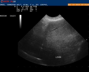
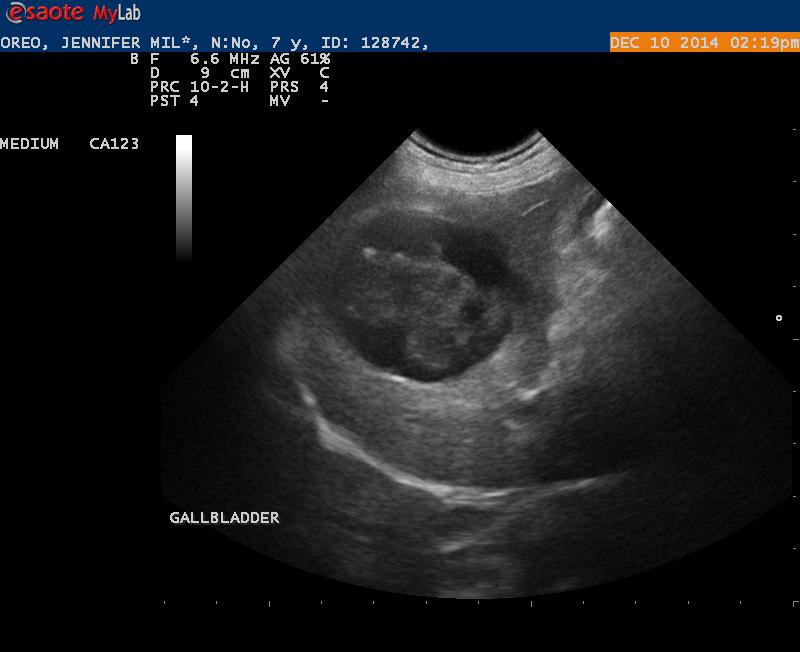
The causes of jaundice would be: pre-hepatic, hepatic and post hepatic. The HCT was normal with no signs of regeneration so that pretty much elmininated pre-hepatic as a cause. When I looked at the liver parenchyma it looked pretty normal. That leaves post hepatic obstruction. I am posting some images and a cine of the gallbaladder and what I consider the dilated CBD.
I have 2 questions:
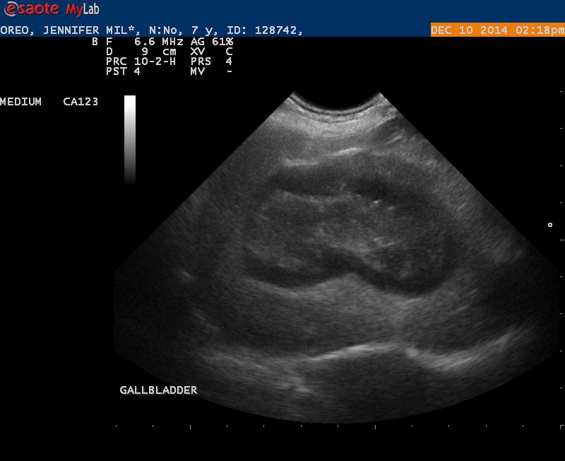
1. Is there an emerging gallbladder mucocoele?
2. Is that a mass I am looking at that is obstructions the CBD?
Thanks for any feedback. This is my first biliary obstructive case.
I have sent Oreo to a surgeon for exploratory. Even if I am wrong with my interpretation here, I am 100% sure there is obstructive disease and Oreo needs to be explored anyway.
Thanks
18 responses to “Post Hepatic Obstruction”
Oreo went to surgery today
Oreo went to surgery today and had the gallbladder removed. Mucocele confirmed. There was dilation of the cystic and CBD but no sign of a tumor or stone obstructing the CBD. Better prognosis for Oreo.
EL,
If you have some time can
EL,
If you have some time can you point out the structures that you are seeing in the cine loop 2.
I am a bit confused about this. I want to make sure what I see is accurate.
Thanks
Hi Randy I had posted on this
Hi Randy I had posted on this but I must have not finalized it. yes classic mucocele and likely induced in part by chronic active pancreatitis EHBO or mucocele or likely both in this case. Good move pulling the trigger. When the right base of the panc is hypoechoiic like that it can tether the cbd… acutely or in chronic active fashion. The GB has the fuzzy fat at 2 oclock on the neck classic for inflamed mucocele they nearly always inflam or rupture in this spot at the neck. Did he have a + Murphy sign in that spot too?
We have a big study on clinical parameters of sx biliary disease (did an abstract in resources) just no time to write it but would like to add your case as well if we can. Check out mucocele in th ebasic search and you will get tired of seeing these presentations 🙂 Its so very very important to recognize them as sx emergencies. Nice job!
Can use my case. Let me know
Can use my case. Let me know if you need any more information or pictures.
Thanks EL,
I believe there
Thanks EL,
I believe there was some leakage. The surgeon saw some adhesion.
Looks like we got to it just in time.
Thanks EL,
I believe there
Thanks EL,
I believe there was some leakage. The surgeon saw some adhesion.
Looks like we got to it just in time.
It likely looked something
It likely looked something like this… direct form another Gbm perf with stones as well sent by a telemed client off the read yesterday. I love when I get outcome from the telemed cases:)
It likely looked something
It likely looked something like this… direct form another Gbm perf with stones as well sent by a telemed client off the read yesterday. I love when I get outcome from the telemed cases:)
Sure Randy in the first image
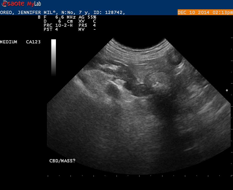
Sure Randy in the first image attached the long arrow points to the dilated cbd > 0.4 cm in a dog is pathological. The small arrow points to the hypoechoic portion of the right panc base and that swallows up the cbd causing EHBO along with other factors like cbd bile sludge infection and so forth.
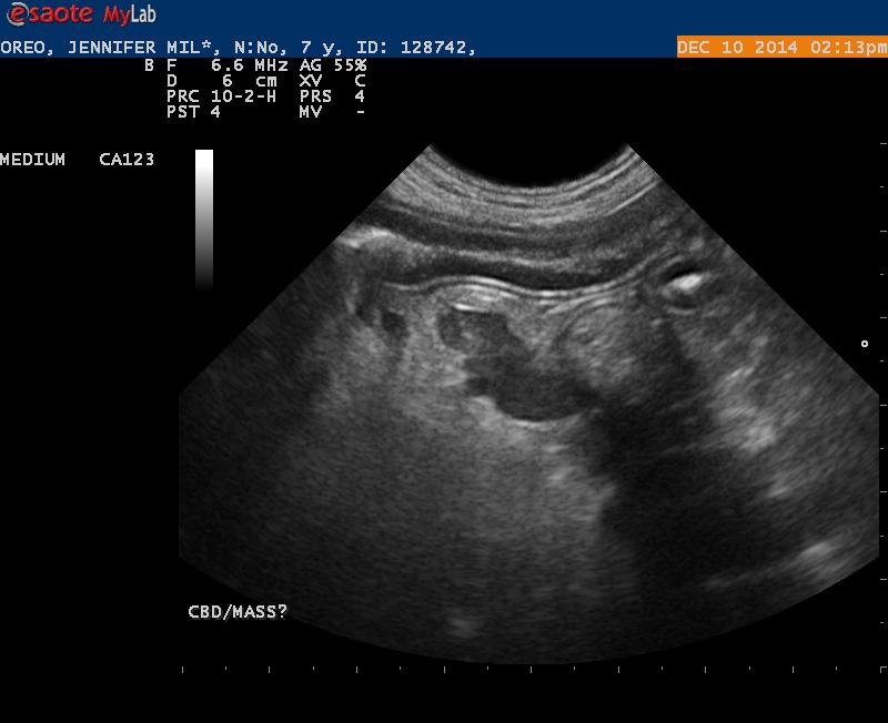
In the second image of the still of your video the neck of the Gb is severly dilated so no conical tear drop shape but now egg shaped and the arrow points to the fuzzy hyperechoic fat that corresponds to inflammation or adhesions and possible perf if local fluid there or perf and absorption if no fluid and Gb collapse. You can see this on the post sx image I posted as well. This is the area you usually get a + Murphy sign unless the patient is really septic and moribund.
Re the pancreas check out this study: ECVIM 2014
and regarding the Sx biliary disease check out this other one we did as well and are continuing to ciollect data for publication: ECVIM 2009 and defining a Gb mucocele survey.
http://sonopath.com/resources/research-publications
I saw the changes in the
I saw the changes in the pancreas and was concerned about pancreatitis- but I was confused about the hypo-echoic region and its relationship to the CBD. You have clarified this for me now. What is the circular shadowing area that is present? Is that gas from the bowel? Its funny how things clarify when someone points it out to you.
Thanks
Sure Randy in the first image
Sure Randy in the first image attached the long arrow points to the dilated cbd > 0.4 cm in a dog is pathological. The small arrow points to the hypoechoic portion of the right panc base and that swallows up the cbd causing EHBO along with other factors like cbd bile sludge infection and so forth.
In the second image of the still of your video the neck of the Gb is severly dilated so no conical tear drop shape but now egg shaped and the arrow points to the fuzzy hyperechoic fat that corresponds to inflammation or adhesions and possible perf if local fluid there or perf and absorption if no fluid and Gb collapse. You can see this on the post sx image I posted as well. This is the area you usually get a + Murphy sign unless the patient is really septic and moribund.
Re the pancreas check out this study: ECVIM 2014
and regarding the Sx biliary disease check out this other one we did as well and are continuing to ciollect data for publication: ECVIM 2009 and defining a Gb mucocele survey.
http://sonopath.com/resources/research-publications
I saw the changes in the
I saw the changes in the pancreas and was concerned about pancreatitis- but I was confused about the hypo-echoic region and its relationship to the CBD. You have clarified this for me now. What is the circular shadowing area that is present? Is that gas from the bowel? Its funny how things clarify when someone points it out to you.
Thanks
The shadow at 2-3 cm int he 4
The shadow at 2-3 cm int he 4 oclock position in the first image I put up there is the ascending/transverse colon with progressively shadowing stool in the lumen.
The shadow at 2-3 cm int he 4
The shadow at 2-3 cm int he 4 oclock position in the first image I put up there is the ascending/transverse colon with progressively shadowing stool in the lumen.