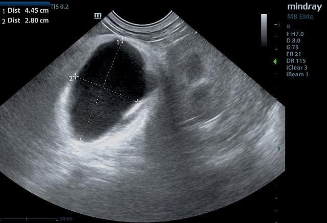
The patient presented for pre-anesthetic work up for a TPLO surgery and it was noted that the patient’s abdomen appeared larger than normal. Radiographs of the abdomen were inconclusive and an ultrasound was performed.
The patient presented for pre-anesthetic work up for a TPLO surgery and it was noted that the patient’s abdomen appeared larger than normal. Radiographs of the abdomen were inconclusive and an ultrasound was performed.