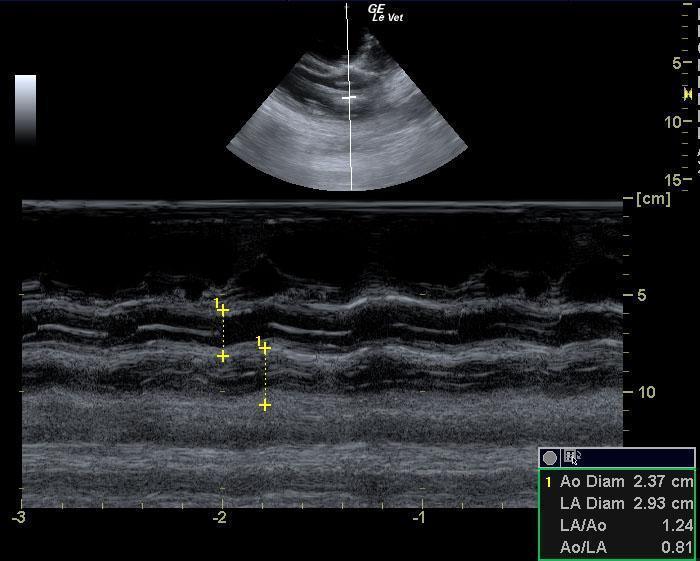
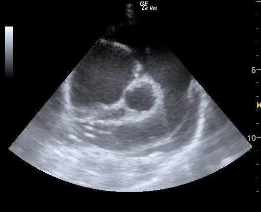
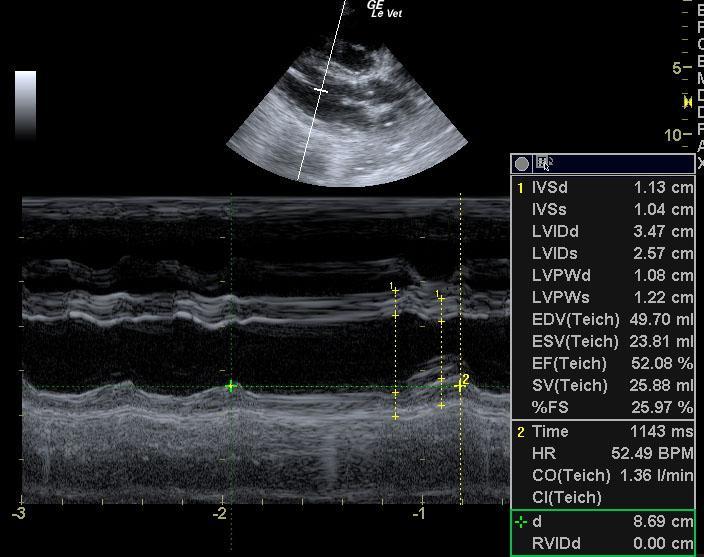
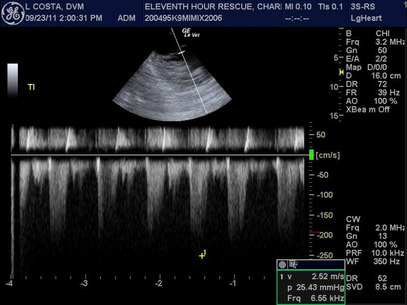
An intact male mixed breed dog was presented for evaluation of 4-5 days of progressive abdominal distension. Abnormalities on laboratory work were heartworm positive and anemia. Survey radiographs showed cardiomegaly and a possible abdominal mass.
An intact male mixed breed dog was presented for evaluation of 4-5 days of progressive abdominal distension. Abnormalities on laboratory work were heartworm positive and anemia. Survey radiographs showed cardiomegaly and a possible abdominal mass.