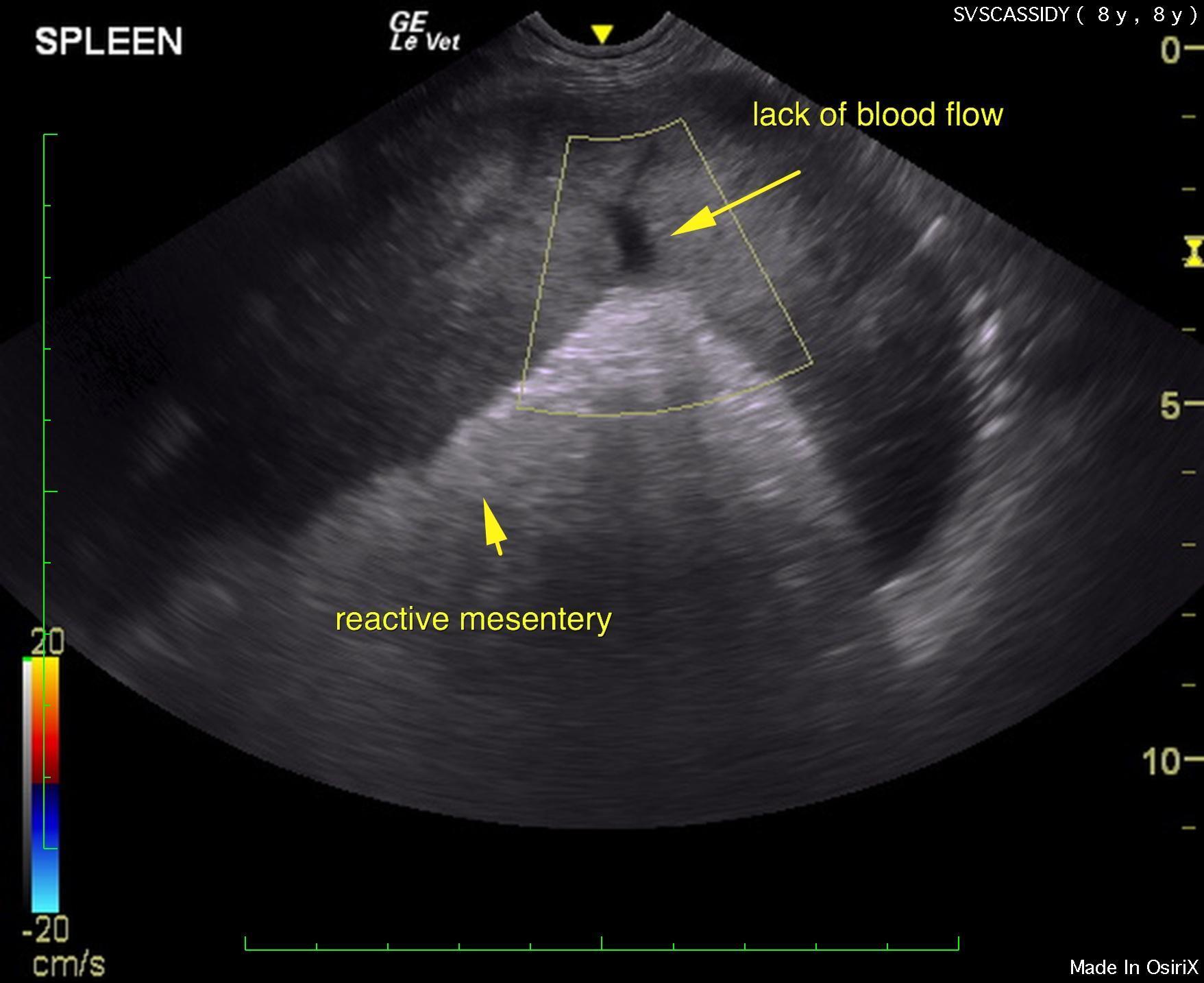
Big spleen, sudden onset, reactive mesentery & nothing else that says neoplasia? Think splenic torsion, remembering the one key button to push is the color flow Doppler over the splenic vasculature. Splenic torsions can look like splenic lymphoma sonographically, but torsions shut off the vasculature. This quick thinking clinical sonographer maneuver will save a life because you don’t let the sun set on a pyometra and you don’t let another minute pass on a splenic torsion. Great work by Sierra Veterinary Specialists & clinical sonographer Loetitia Saint-Jacques RVT, LVT of Paws Mobile Sonography, Lake Tahoe, CA, USA.
The patient was presented on emergency from his RDVM. Thoracic radiographs were unremarkable. 2-view abdominal radiographs showed a cranial abdominal mass. CBC: HCT 32%, Anemia, elevated WBC 29.58, Mono 59, Neut 22.44, PCV/TP 36%/8.2 upon presentation. A double cavity ultrasound was performed.
The patient was presented on emergency from his RDVM. Thoracic radiographs were unremarkable. 2-view abdominal radiographs showed a cranial abdominal mass. CBC: HCT 32%, Anemia, elevated WBC 29.58, Mono 59, Neut 22.44, PCV/TP 36%/8.2 upon presentation. A double cavity ultrasound was performed.
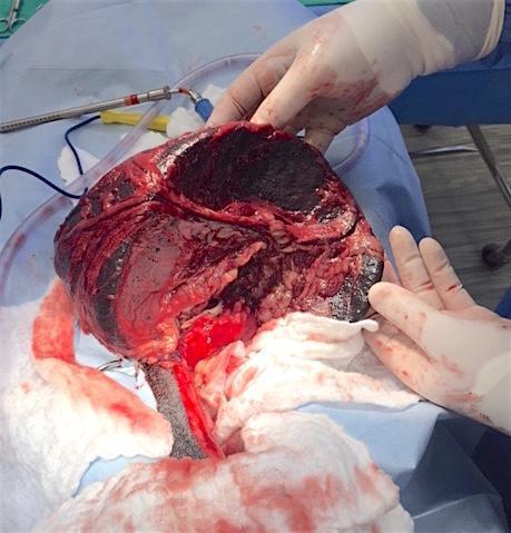
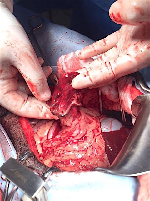
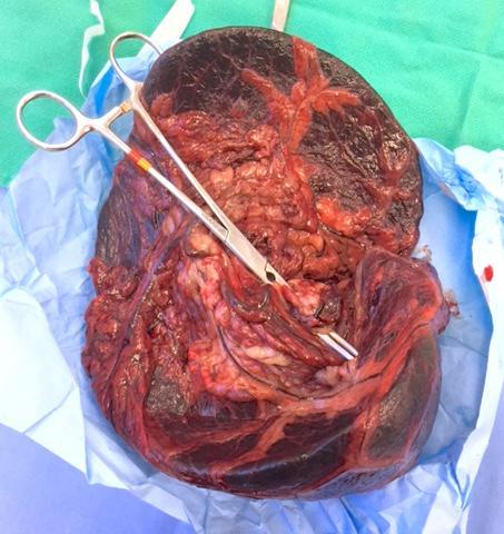
Acute splenic torsion with regional peritonitis.
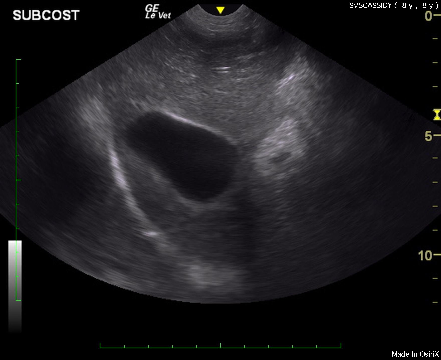
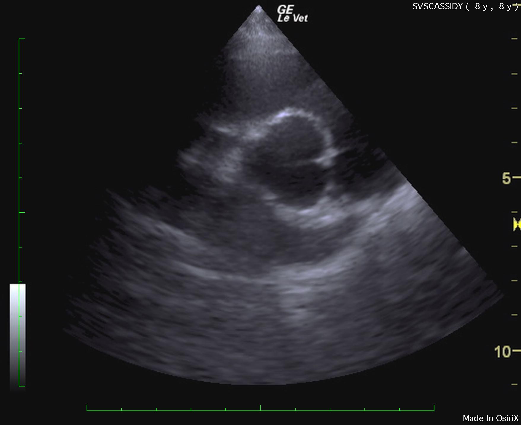
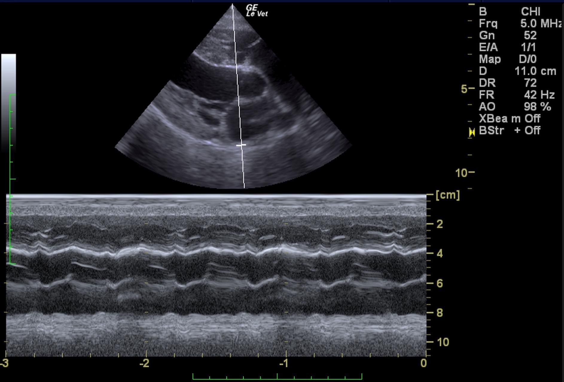
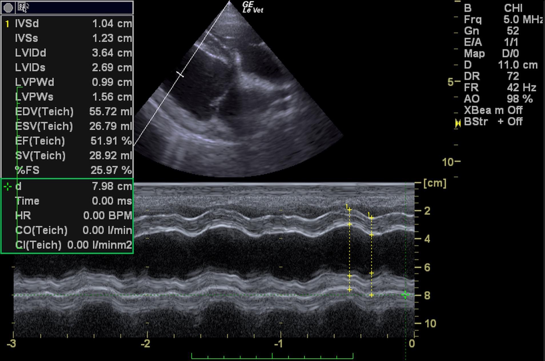
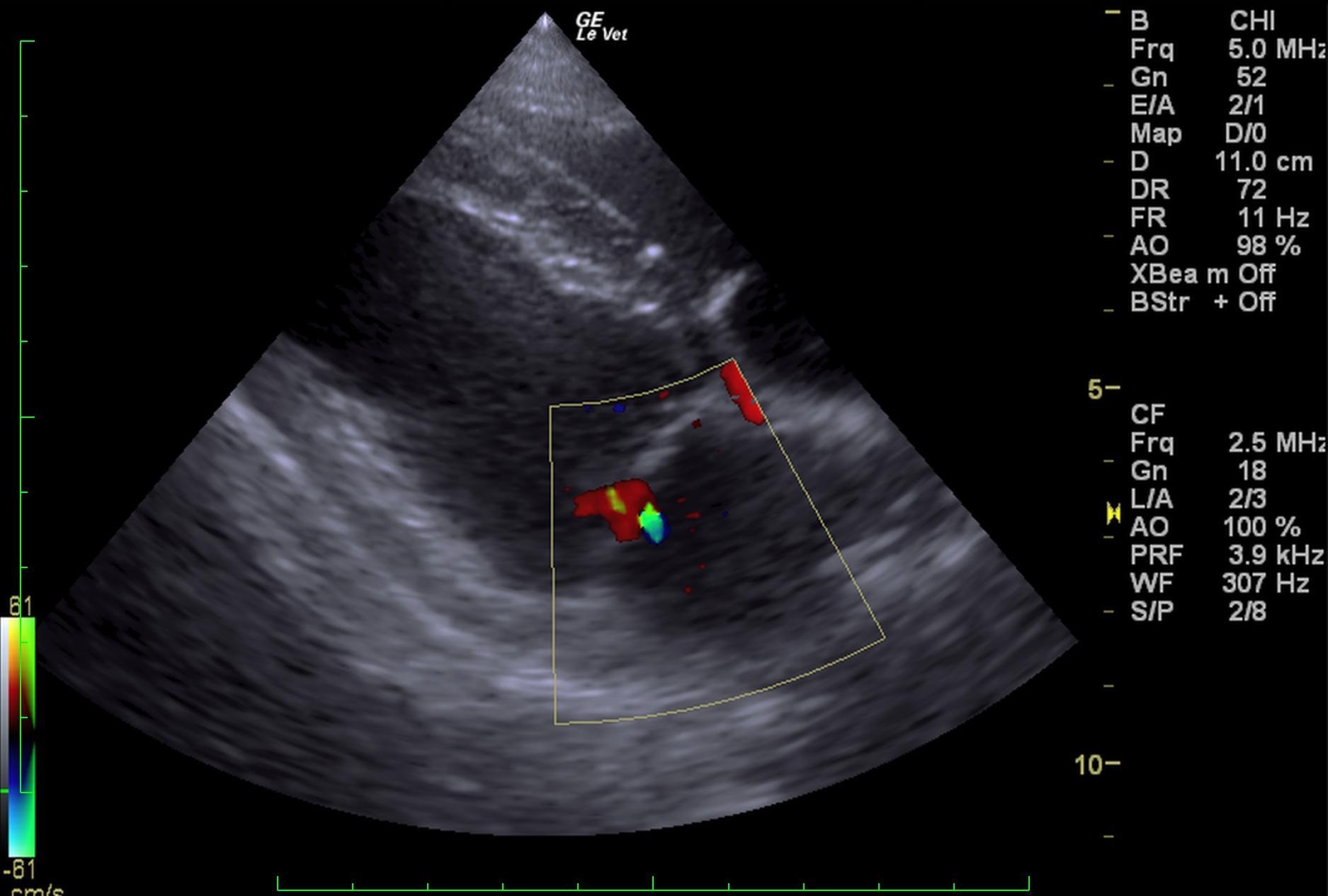
The spleen in this patient revealed severe enlargement and reactive mesentery. Irregular contour was noted as well as micronodular changes that extended throughout the abdomen. This is strongly consistent with splenic torsion and appeared to be isolated to the spleen. A slight amount of free fluid was noted in the caudal abdomen adjacent to the bladder. Reactive mesentery was noted throughout the mid-abdomen. The liver was slightly swollen, yet not obviously enlarged in the pathological process. Normal echocardiogram with trivial mitral insufficiency.
Immediate exploratory surgery was recommended. Underlying neoplasia is possible, yet unlikely. A splenectomy was performed. No contraindication to anesthetic procedure based on echocardiogram of normal volume, function and lack of right auricular masses or pericardial effusion.