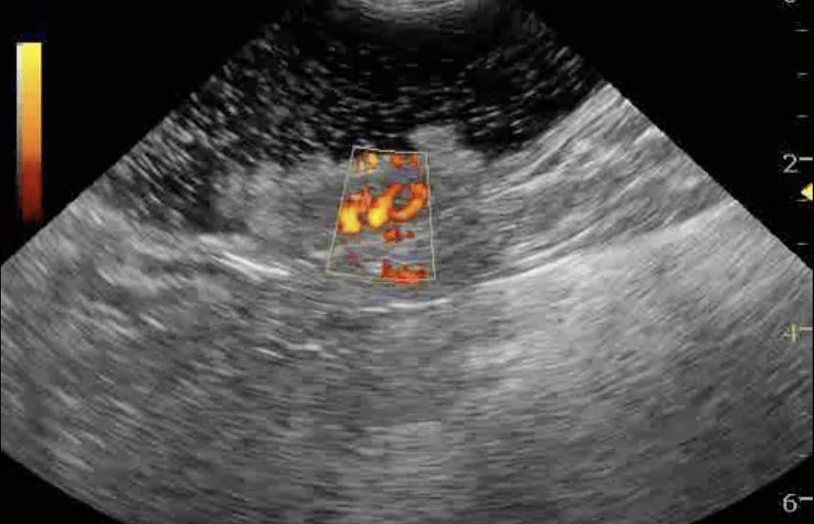
This is an ultrasound-guided endoscopic diode laser ablation (UGELAB) of a transitional cell carcinoma in the uirinary bladder of a 10-year-old FS Beagle. There were many polyps in the trigone and urethra that were ablated as well. Rosie is doing well 12 months post UGELAB. This procedure was performed by Dr Dean Cerf (Laser/scope) and Dr Eric Lindquist at Dr. Cerf’s facility of Ridgewood Veterinary Hospital, Ridgewood, NJ, USA.
This is an ultrasound-guided endoscopic diode laser ablation (UGELAB) of a transitional cell carcinoma in the uirinary bladder of a 10-year-old FS Beagle. There were many polyps in the trigone and urethra that were ablated as well. Rosie is doing well 12 months post UGELAB. This procedure was performed by Dr Dean Cerf (Laser/scope) and Dr Eric Lindquist at Dr. Cerf’s facility of Ridgewood Veterinary Hospital, Ridgewood, NJ, USA. The original abstract for this procedure was presented at ACVIM (Seattle)and ECVIM (Budapest) 2007 and will be also presented for a full hour lecture at ACVIM June 5, 2008 in San Antonio, Tx.
UGELAB (TCC)
History
Comments
After consultation with the pathologist, the hypertrophy of the muscularis was physiological in response to functional obstruction or a “pseudohypertrophy syndrome.” Only islands of cross over infiltrative lymphoma were visualized within the submucosae and muscularis while the diagnosis was consistent with diffuse, low grade mucosal lymphoma typical of older cats. In his opinion, these patients tend to respond better to therapy and have a less aggressive form of lymphoma than the forms that involve the submucosal and muscularis layers in a diffuse manner. Since the regions of detail loss of the intestinal layers were minimal, physiological hypertrophy of the muscularis is most consistent with the sonographic presentation as opposed to infiltrative disease of the muscularis/submucosae. The minor areas of detail loss between the muscularis, submucosae and mucosae may have corresponded to the “islands” of cross-over infiltrate that the pathologist had described (see linear intraop. image 312L). Concurrent mast cell disease of the mesenteric lymph node was considered a complicating factor but would most likely not be treated differently. Regardless, the owner declined aggressive chemotherapy. The patient was prescribed an oral, bland chemotherapeutic protocol of immune suppressive prednisolone, chlorambucil and broad spectrum antibiotics. The cat responded well to therapy, gaining weight with a strong appetite 8 months post diagnosis.
Referring Practitioner and diode laser operator: Dean Cerf DVM, Ridgewood Veterinary Hospital, Ridgewood, NJ, USA.
Sampling
Laparotomy was performed with intraoperative imaging of the intestinal tract in order to isolate the small intestinal thickening of the muscularis. A focal region of intestinal perforation was present without significant serosal changes and minor surgically palpable thickening of the segment in question. The surgeon resected of the portion of the bowel that contained thickened muscularis according to the intraoperative sonographer’s direction. Full thickness biopsy of the small intestine with normal muscularis thickness was also obtained as well as mesenteric lymph node and liver. Samples were placed in separate biopsy jars and labeled accordingly as to the sonographic presentation. Histopath: Intestine with muscularis thickening: First read was interpreted as intramural lymphocytic enteritis with muscularis hypertrophy. Second opinion interpretation: Low grade, diffuse, mucosal intestinal lymphoma with muscularis hypertrophy and mild multifocal submucosal and muscularis lymphoid infiltrate. Lymph node: low grade mast cell neoplasia. Liver: Moderate chronic lymphocytic hepatitis.
Sonographic Differential Diagnosis
Differential Diagnosis: Intestinal lymphoma, mast cell disease,IBD, FIP, carcinoma less likely.
Video
Patient Information
Images