Teamwork makes the dreamwork! Team scanning is a fantastic way to learn other’s imaging techniques, tips, and tricks. In this Case Of the Month, we are focused on “old cat tubes”, imaging of the common bile duct, duodenal papilla, and biliary tree. This 13-year-old cat had some pathology plugging up the tubes. See what our team found with images provided by Shari Reffi, CVT, SDEP® Certified clinical sonographer for SonoPath Mobile Veterinary Ultrasound and Jessica Miller, BS, RDMS, clinical sonographer for SonoPath Mobile Veterinary Ultrasound. Interpretation of ultrasound images by Eric Lindquist, DMV, DABVP, Cert. IVUSS and cytology interpretation by L.D. McGill, DVM, Ph.D., DACVP.
Decreased appetite. Current meds: Metronidazole, Denamarin, Mirtazapine. ALT 393, T. bili 5.7, Mag 2.7, Chol 307, Amyl 1891, PSL 50, Lymphs 9, Mono 8, Neuts 11,680, Mono 1280,Eos. 1600, USG 1.048.
Decreased appetite. Current meds: Metronidazole, Denamarin, Mirtazapine. ALT 393, T. bili 5.7, Mag 2.7, Chol 307, Amyl 1891, PSL 50, Lymphs 9, Mono 8, Neuts 11,680, Mono 1280,Eos. 1600, USG 1.048.
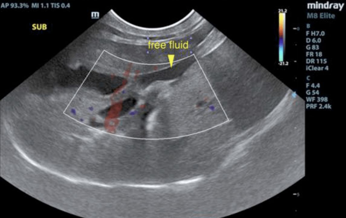
Hepatic swelling with common bile duct dilation and free fluid. Biliary calculi, may be incidental. Scalloping spleen.
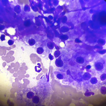
Spleen: The spleen was mildly enlarged with uniform, but subtly micronodular parenchyma, and undulating capsular contour. This is consistent with reactive spleen owing to immune stimulus or early infiltrative disease such as mast cell disease or lymphoma.
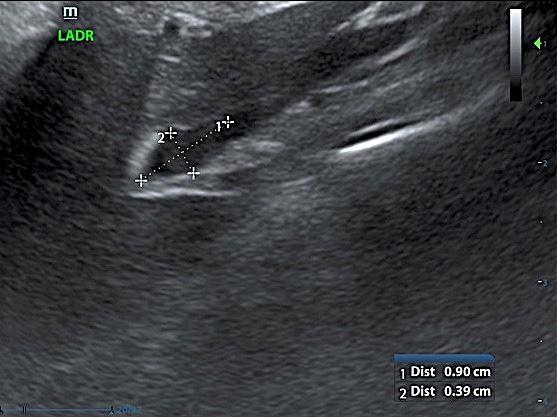
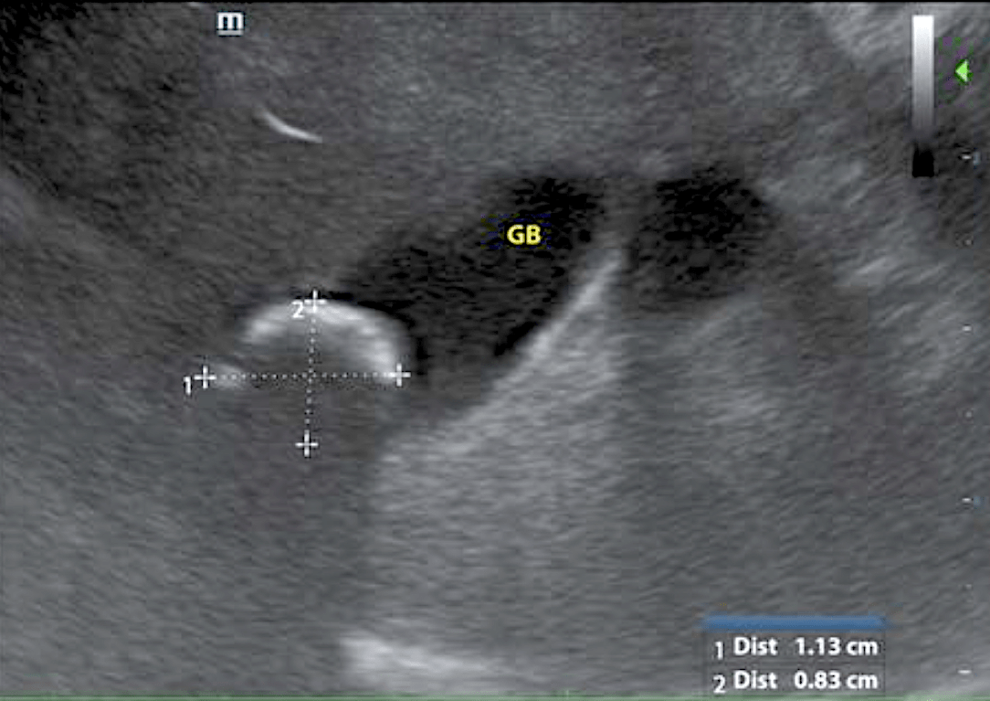
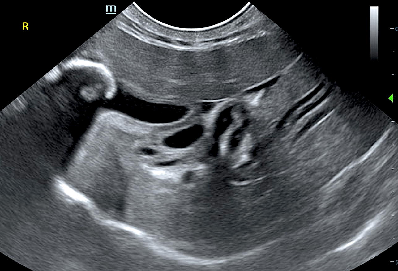
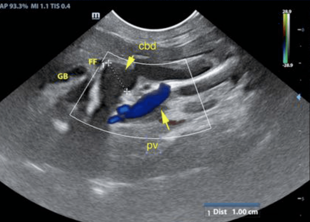
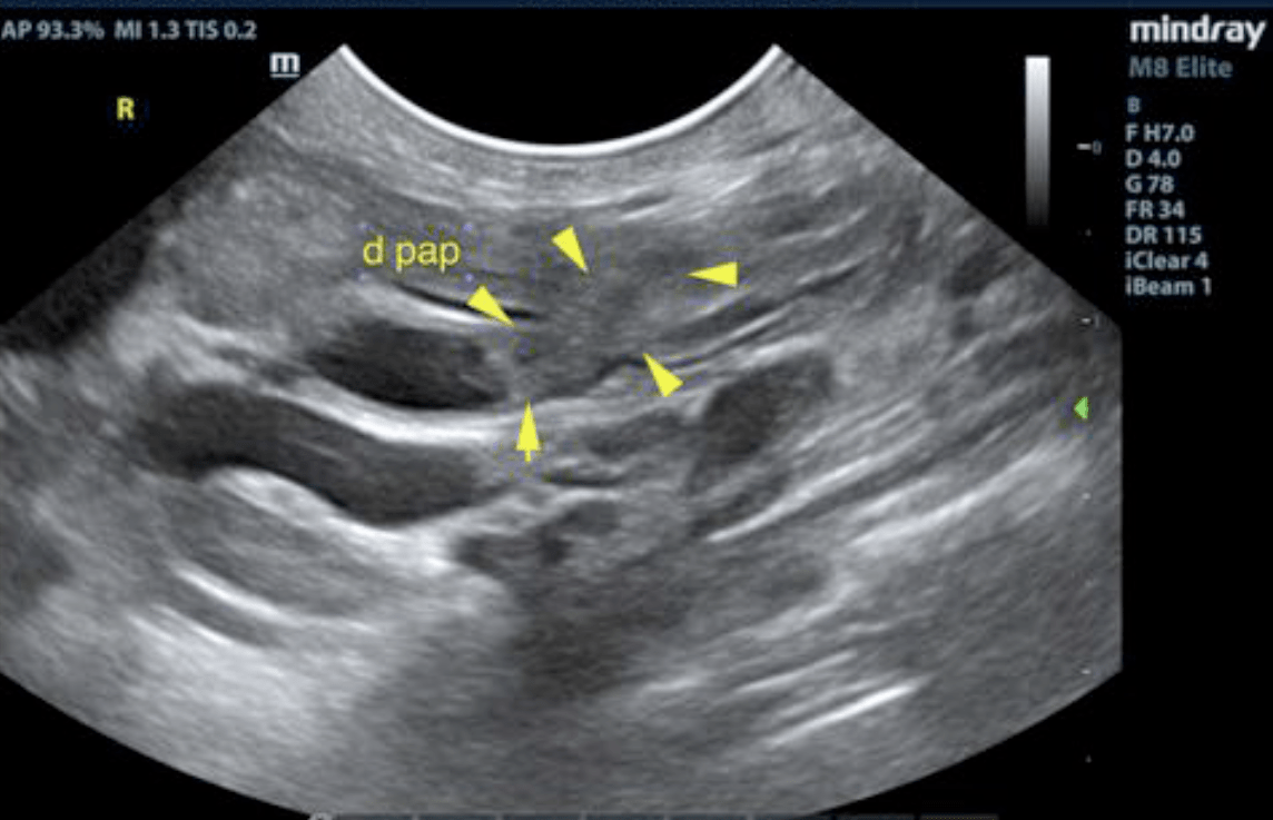
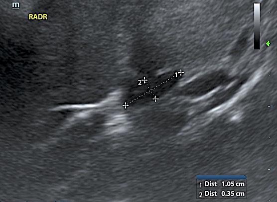
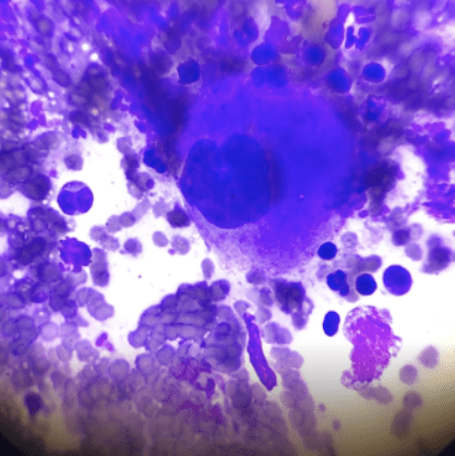
Liver: The hepatic parenchyma was uniformly swollen. Lobar biliary duct dilation was noted owing to post hepatic obstruction. The gallbladder revealed a 1.13 cm calculus with echogenic debris. The gallbladder wall was mildly thickened and echogenic. The common bile duct was followed to the duodenal papilla. The common bile duct appeared to taper normally, yet the common bile duct was excessively dilated up to 0.5 cm. The hepatic lymph nodes are slightly enlarged and measured 0.6 cm.