Head to Toe Diagnostic imaging. When the shunt hunt hits a snag sonographically then look to CT for the answer. “After a frustrating 45 minutes of shunt hunting I finally, reluctantly, deferred to CT to further define this vascular anomaly…defeated and down, dragging my probe out the clinic, grumbling as I loaded up for the next mobile appointment in semi defeat. Just then I thought “when ya can’t beat em, join em”… so SonoPath Teleradiology interpreted the CT …and then SonoPath bought one.” -Dr. Eric Lindquist 🙂
Portophrenic shunt? What the heck is that? Well, leave it to a Boxer to throw a congenital curve at ya.
A big thanks to the following teams of people for their many contributions to the management of this difficult case. Animal Clinic of Morris Plains, Advanced Veterinary Care, Anjilla Cooley DVM MS. Small Animal Surgery, SonoPath Mobile Veterinary Ultrasound, SonoPath Teleradiology. We needed a community to solve this Boxer mystery.
The patient was presented for several days of lethargy, PU/PD, inappetence, but no vomiting. Numerous congenital musculoskeletal abnormalities were apparent. Blood chemistry found globulins 4, ALT 548, all else WNL.
The patient was presented for several days of lethargy, PU/PD, inappetence, but no vomiting. Numerous congenital musculoskeletal abnormalities were apparent. Blood chemistry found globulins 4, ALT 548, all else WNL.
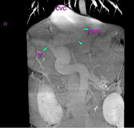
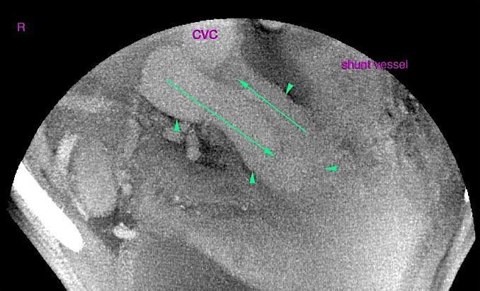
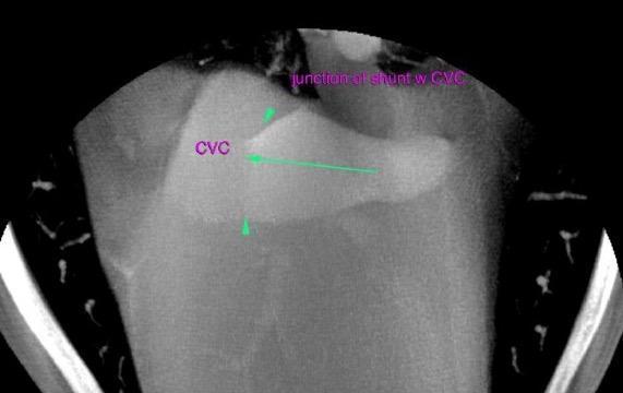
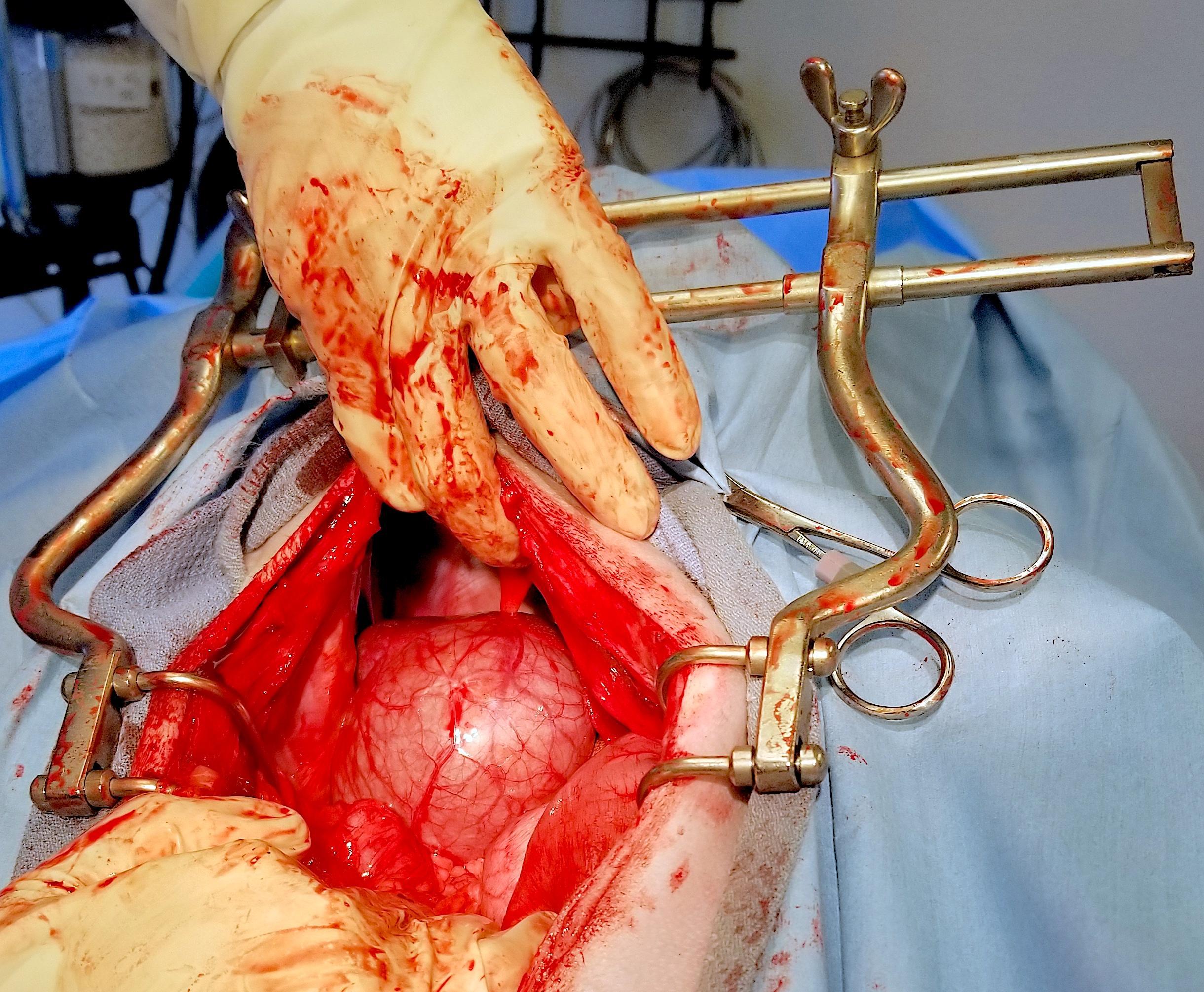
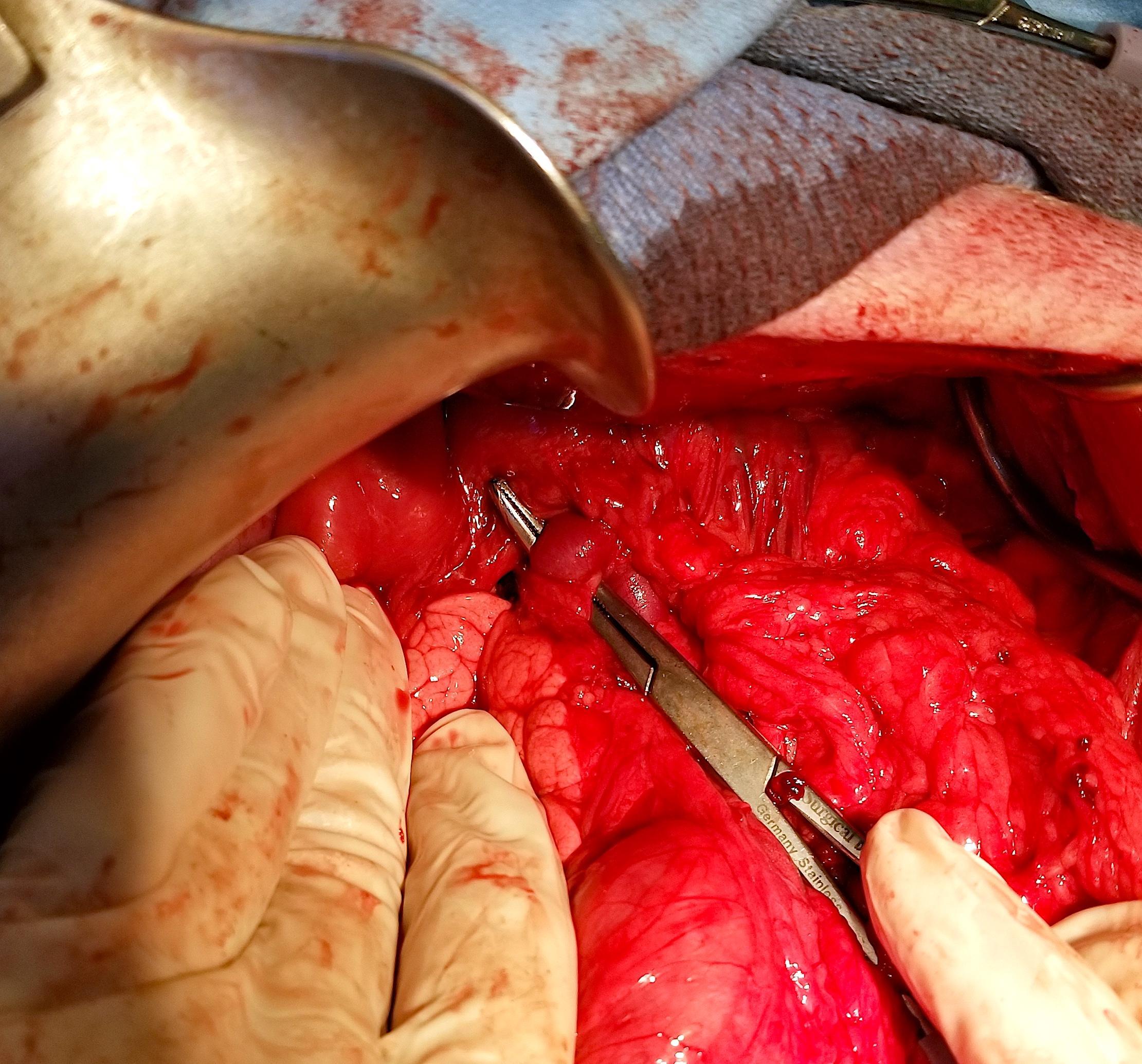
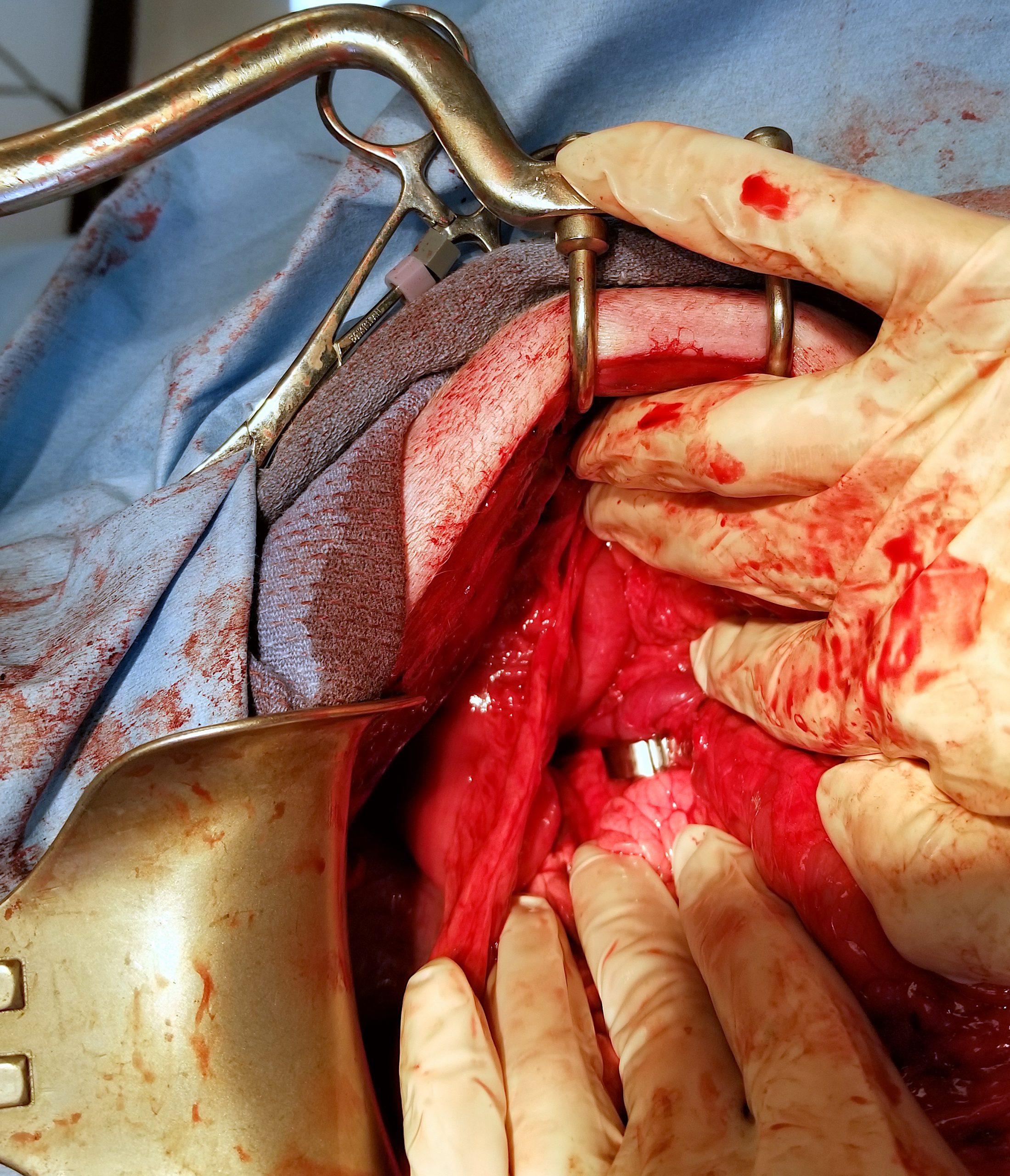
U/S: Liver shunt requiring further definition. Final CT diagnosis was a single congenital extrahepatic portosystemic shunt of the porto-phrenico type.
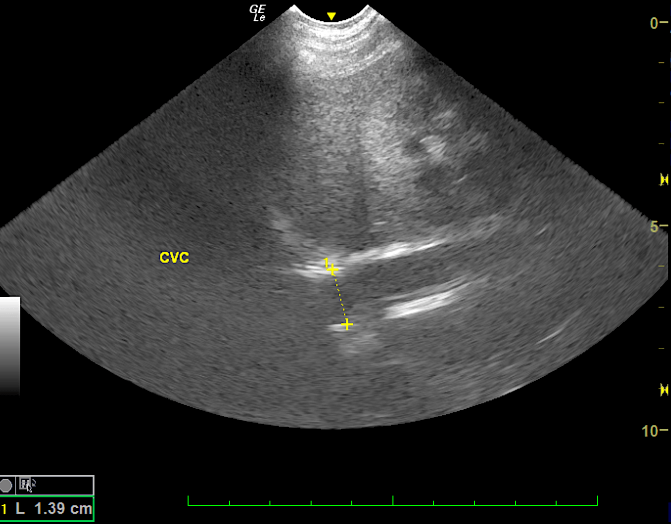
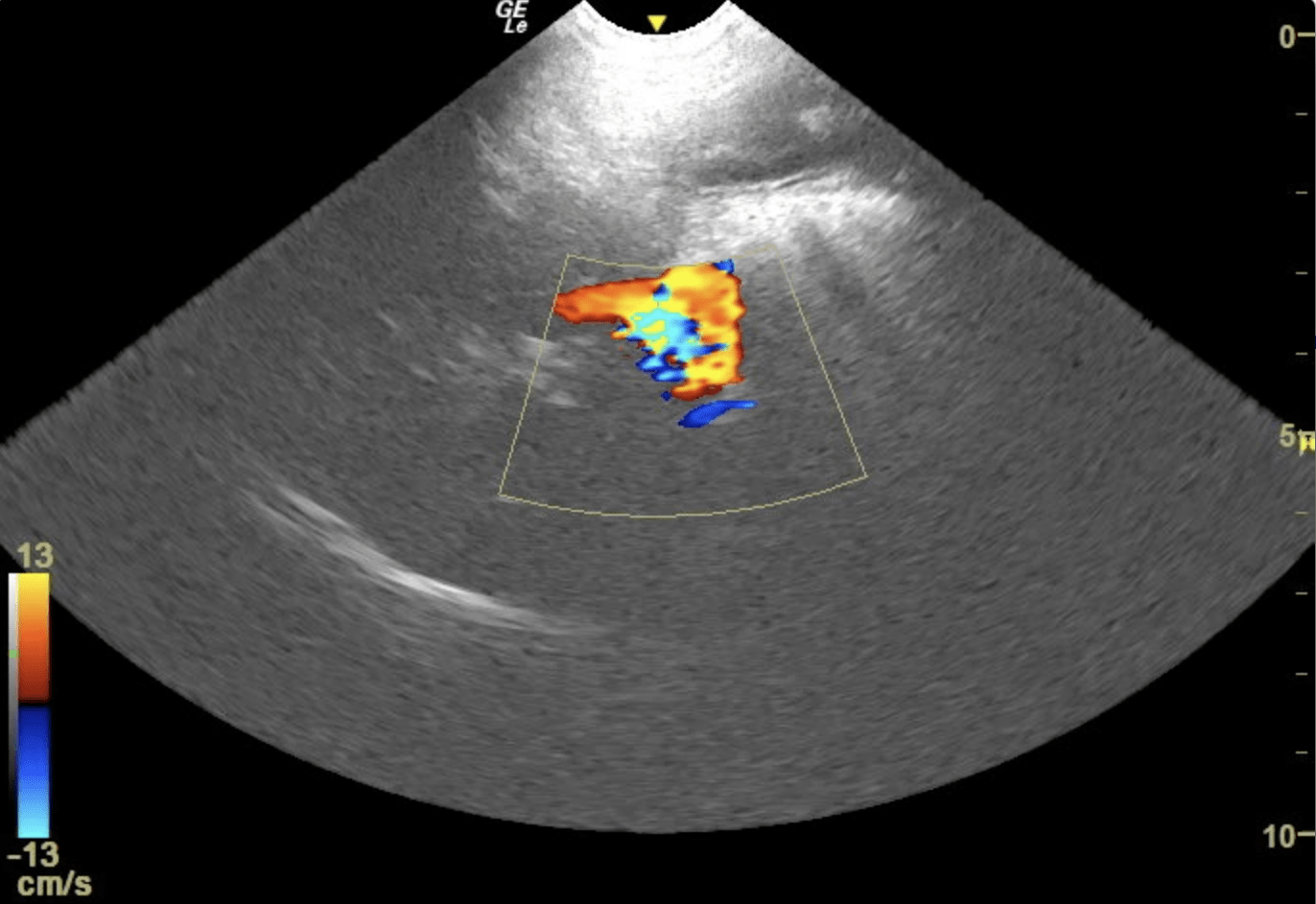
The liver demonstrated intrahepatic hypovascularity and overwhelming gallbladder dilation, yet this is not to the level of mucocele formation. Acoustic windows were extremely limited owing to the confirmation of this patient. However, portosystemic shunt was found at the level of the portal hilus, yet appeared to be in the position of a short, right divisional intrahepatic shunt. However, extrahepatic shunt could not be excluded as the visibility was extremely poor and views of the actual anomalous vessel were limited, not allowing for differentiation of intrahepatic or extrahepatic position. The shunt measured 1.2 cm wide.
CT evaluation was recommended for surgical planning to evaluate for both the possibility of extrahepatic and intrahepatic shunt. Given the breed and position of the shunt, it would be most consistent with right divisional intrahepatic shunt, yet should be further evaluated by CT. In the meantime, given the confirmed elevated bile acids, medical treatment was recommended for this patient until further intervention could occur. CT evaluation found a single abnormal extrahepatic vessel connecting the portal vasculature with the systemic circulation. The dilated shunt vessel emerged from the portal vein level with the junction of the splenic vein, bypassed the liver dorsally and joined the caudal vena cava at the diaphragm. The extrahepatic portal vein was uneven and small in diameter. The hepatic artery presented compensatory enlargement. The intrahepatic branching of the portal vein was reduced. Second order portal veins could be seen within the hepatic parenchyma. Mild microhepatica was noted. CT was able to identify a congenital extrahepatic porto-phrenic shunt which commonly gets mistaken for an intrahepatic shunt.