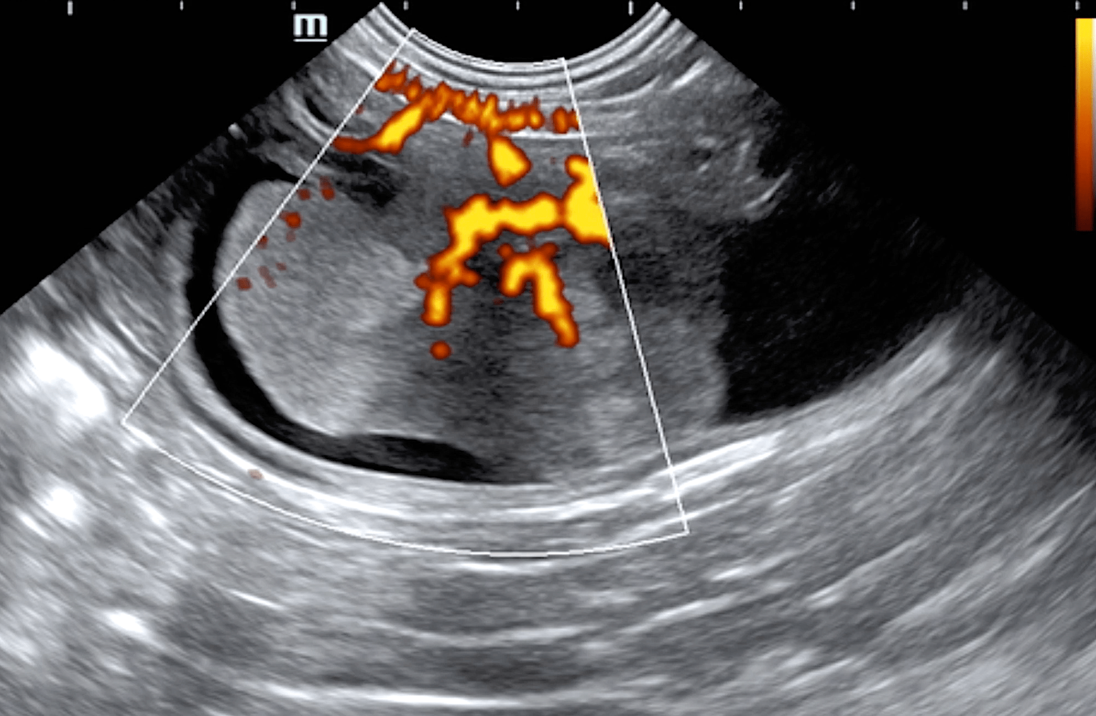
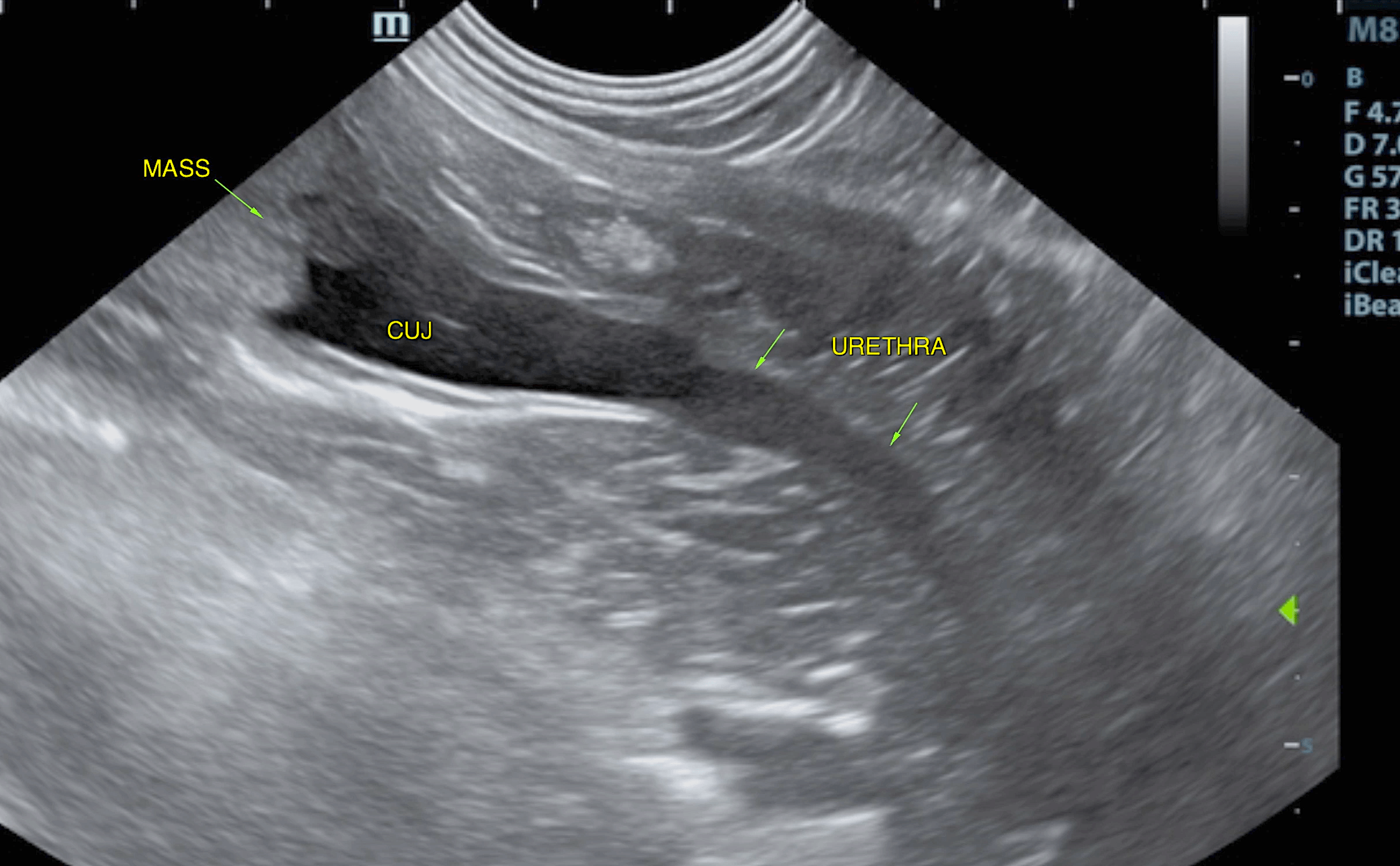
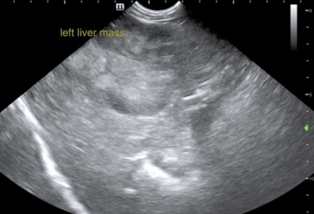
Presented with clinical signs of a UTI or worse? Schedule an ultrasound earlier rather than later especially if nonresponsive to a first round of treatment. When you see a mass like this, think – Is it resectable and how do I evaluate for resectability? We’ve got you covered with key SDEP positions 1-3: long axis fan, short axis fan, Doppler of the mass, long and short axis of the cysto-urethral junction, and a long urethra into the pelvis. We recommend at least 4cm if not more, to fully evaluate for any masses further down the road and avoid surprises later. Decrease your depth to eliminate wasted space under the bladder, bring out that linear probe and focus on the ureters to look for possible ureteral invasion (see our Case of the Month July 2018). And don’t be tempted to only assess the primary issue – always do a full abdominal scan, so as not to miss other pathology such as the liver masses in this case. These brilliant images are by one SonoPath’s own Diane McFadden, RVT, SDEP™ Certified Clinical Sonographer, Director of Mobile Operations and Education. Special thanks to Dr Lee, Dr Logan and the staff at Mount Olive Veterinary Hospital for their management of this case in providing an answer and options for the owners.
The patient was presented due to panting, urinary accidents, PU/PD, +1 polyphagia, hepatomegaly on radiographs. Urinalysis revealed hematuria, pyuria, and hyposthenuria. U/A: USG 1.002, protein +2, WBCs 4-10, RBCs 11-20, rods 26-50. Blood chemistry results: ALT 283, Alk. Phos. 226.
The patient was presented due to panting, urinary accidents, PU/PD, +1 polyphagia, hepatomegaly on radiographs. Urinalysis revealed hematuria, pyuria, and hyposthenuria. U/A: USG 1.002, protein +2, WBCs 4-10, RBCs 11-20, rods 26-50. Blood chemistry results: ALT 283, Alk. Phos. 226.
Bladder mass. Left-sided liver masses.
Urinary bladder: revealed a ventral bladder mass that measured 3.0 x 2.5 cm. The mass was deriving from the ventral wall and extended caudally along the ventral wall to approximately 1.0 cm from the cystourethral junction. This allowed for resection. A minimal amount of urine was present and impinged upon the dorsal wall. The mass was particularly vascular. This is typical for transitional cell carcinoma. This is potentially resectable. However, local metastasis to the dorsal wall with a kissing lesion cannot be entirely ruled out as a minimal amount of urine was present at the time of the sonogram. Cystourethral junction and urethra appeared unremarkable. The pelvic urethra was imaged 4.0 cm beyond the cystourethral junction.
Liver: presented multiple masses, which is likely independent from the bladder mass and impinged upon the portal hilus and gallbladder. Subtle, heterogenous right-sided changes were noted. CT evaluation is warranted for surgical planning.


