What causes an expanding set of kidneys in an “ADR” middle-aged Beagle in renal failure? Holy hematocrit Batman! Is that erythropoietin gone awry? Put a probe on it and a needle in it and see what comes up. Its a matter of point and shoot for needle slinger Dr. Doug Casey DABVP of English Bay Ultrasound, Vancouver BC, Canada in the May 2014 case of the month.
A 7-year-old FS Beagle was presented for vomiting, lethargy, and anorexia. Blood chemistry showed a high creatinine, hypercalcemia (not ionized), hypoalbuminemia, and hyperglobulinemia. CBC showed polycythemia with a hematocrit of 65%.
A 7-year-old FS Beagle was presented for vomiting, lethargy, and anorexia. Blood chemistry showed a high creatinine, hypercalcemia (not ionized), hypoalbuminemia, and hyperglobulinemia. CBC showed polycythemia with a hematocrit of 65%.
Renal, chronic kidney disease, pyelonephritis, neoplasia. Hypercalcemia, neoplasia, granulomatous desease, hyperparathyroidism, renal disease. Polycythemia, vera, secondary (pulmonary disease, erythropoietin producing tumor).
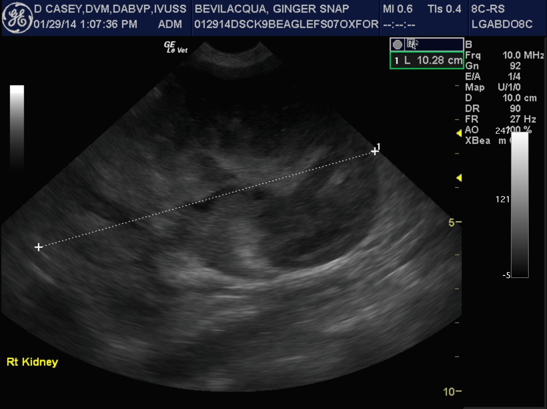
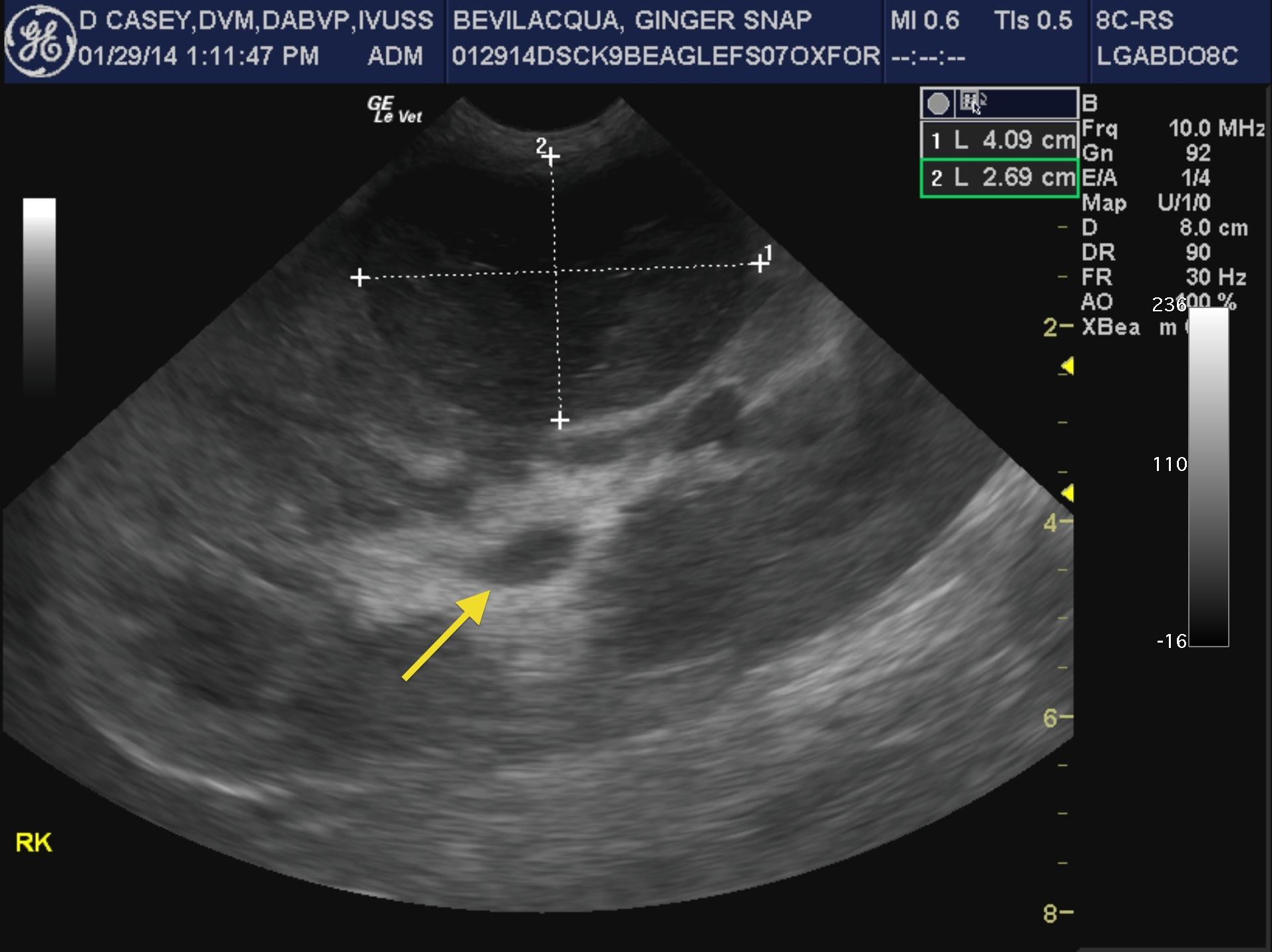
The right kidney in this patient presented a mixed, hypoechoic mass that measured 10.4 x 8.08 cm with hyperechoic surrounding fat. The right kidney was completely infiltrated with a separate mass that measured 4 x 2.69 cm. Pyelectasia was noted in the right kidney and measured 1.08 cm. The right kidney measured 10.28 cm. Complete disruption of the corticomedullary junction and renal pelvis was noted. The left kidney was also enlarged with mild degenerative changes and pyelectasia that measured 0.5 x 1.5 cm. The left kidney measured 5.11 cm.
Ultrasound-guided FNA revealed renal lymphoma The patient was somewhat stable on CCNU and prednisone therapy months after diagnosis of renal lymphoma.
Erythropoietin levels were not taken in this patient however EPO hypersecretion was suspected given the clinical presentation.