Sometimes life’s a beach! This young and playful Golden Retriever was presented with unilateral nasal hemorrhage. CT was able to demonstrate focal destruction of the nasal turbinates by foreign material which was removed after the CT diagnosis and turned out to be a souvenir from his last trip to the beach. Thank you to Nele Ondreka, DVM, Dr. med. vet., DipECVDI for her interpretation of this interesting case and to Dr. Meaux of Mobile Pet Imaging for providing these diagnostic CT images.
A 5-year-old, FS, Golden Retriever was presented for right-sided epistaxis. A pre and post contrast CT of the patient’s head was submitted for interpretation.
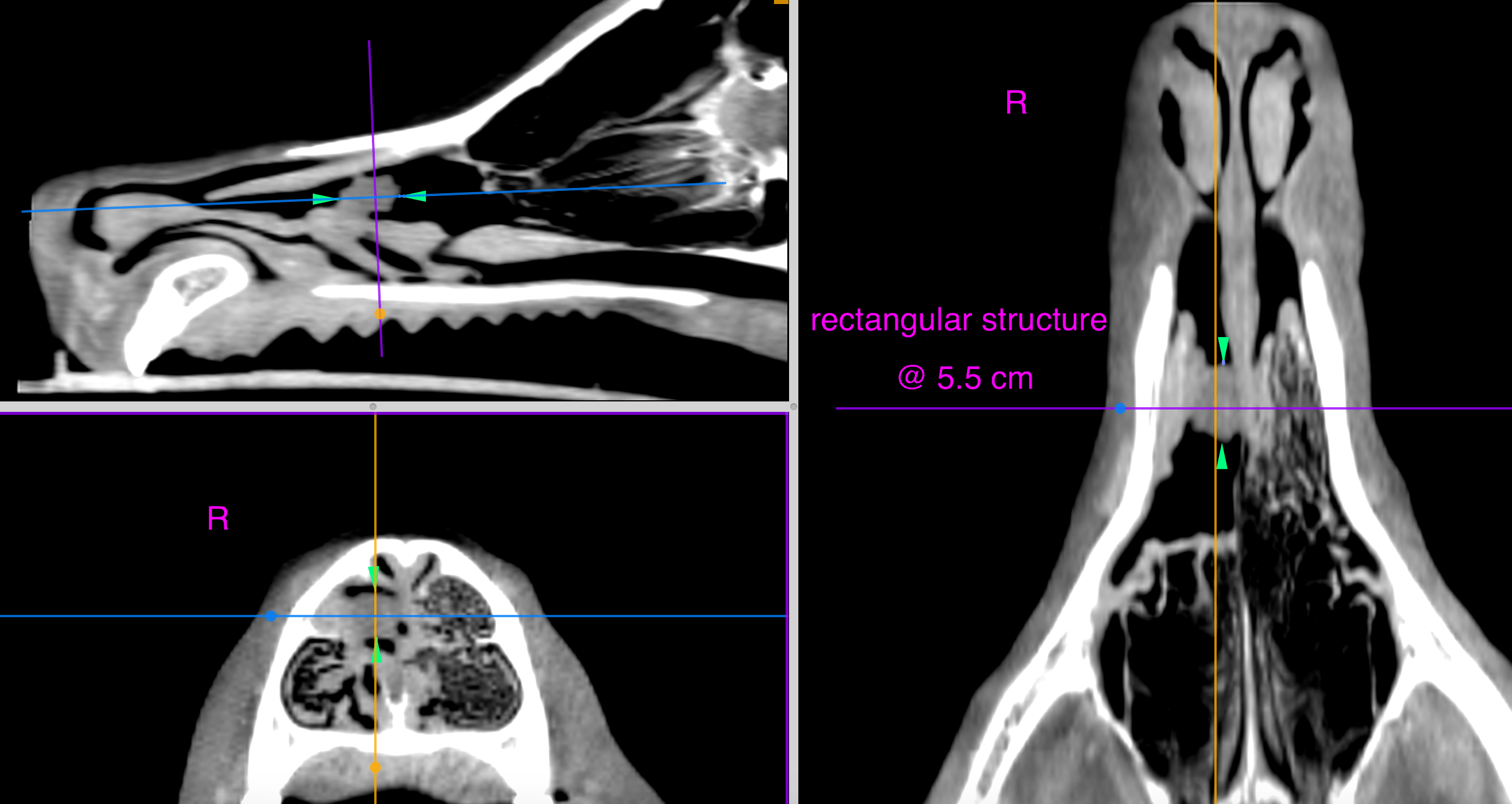
A 5-year-old, FS, Golden Retriever was presented for right-sided epistaxis. A pre and post contrast CT of the patient’s head was submitted for interpretation.
Probable foreign object within the right nasal cavity with chronic, regionally destructive rhinitis/conchal necrosis within right nasal cavity and mild extension into the left nasal cavity through the nasal septum.
A rectangular hypoattenuating structure of 10 mm length and 6 mm height and width, respectively, is seen within the mid third of the right nasal cavity. The structure is located in the position of the former middle nasal meatus and is negative for contrast enhancement. Marked regional turbinate destruction is noted, as well as focal destruction of the nasal septum, adjacent to the rectangular object with mild extension of the destructive changes into the left nasal cavity. Slight regional mucosal swelling is noted, as well as a mild amount of fluid attenuating material on the floor of the right nasal cavity. The submandibular and medial retropharyngeal lymph nodes present within normal limits.
The CT findings were suggestive for foreign body related regional destructive rhinitis. Superinfection could not be ruled out even though the changes seem to be restricted to the region of the probable foreign object. The attenuation pattern was compatible with organic material. Rhinoscopy with removal of the probable foreign object and sampling for culture and sensitivity was recommended. A cluster of sand and a small piece of a shell were retrieved from the right nasal passage via red rubber catheter flush and the patient is doing well post-procedure.