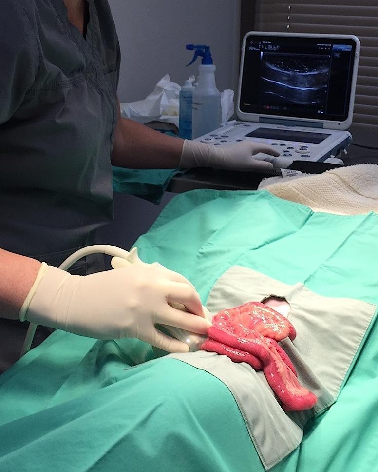
“When I started doing intraoperative ultrasound IOP-US in New Jersey in 2003, stimulated by knowing the lesion is more dramatic than the surgical biopsies resulted to be over and over again, we found the IOP-US applications to be vast and precise to obtain the most representative lesion through the sonogram, evidenced in our mini abstract at ECVIM a few years ago (Intra-operative ultrasound). IOP-US allows us not to play the surgical biopsy guessing game of sampling simply based on the superficial serosal appearance on the organ to be sampled, whether GI, liver, pancreas, kidney,,,,. This case of the month is a bit different than we have done in the past. We made a live typical SonoPath member forum post, provided by long time SonoPath member Jacquie Pankatz DVM, where she applied intraoperative ultrasound to this protein losing enteropathy (PLE) case in order to obtain the perfect histopath representative of the lesion. IOP-US is easy to do. You have a scalpel and a probe so why not put them together and enhance your sampling precision? The pet owner will think you are a genious… and likely some of your staff too. This is a sonographic “mojo” call to the probe. 🙂 Nice job Jaquie! You’ve got game!”, Dr. Eric Lindquist.
A 6-year FS Havanese with history of vomiting (mostly at night and in morning) for a few weeks; no diarrhea. Recent bloodwork found ALB 16, TP 40, GLOB 24 with other parmaters WNL. A fecal was negative. Urine SG 1.035, no protein on chemstrip and urine pro:crea ratio was pending.
A 6-year FS Havanese with history of vomiting (mostly at night and in morning) for a few weeks; no diarrhea. Recent bloodwork found ALB 16, TP 40, GLOB 24 with other parmaters WNL. A fecal was negative. Urine SG 1.035, no protein on chemstrip and urine pro:crea ratio was pending.
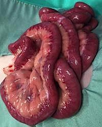
Severe primary Lymphangiectasia.
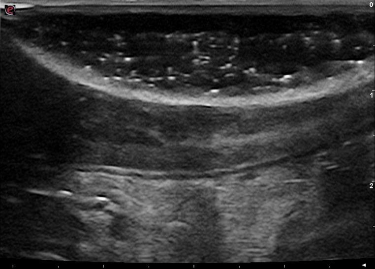
Ultrasound showed an approx. 4cm length of small intestine with a uniformly ill-defined hyperechoic mucosal layer (“mucosal fogging”-EL) with overall normal wall thickness and normal layering; the classic mucosal striations were not seen, but some mucosal speckling was present. The more dramatic lesion (fogging) was segmental rednering IOP-US ideal to delineate and sample.
Dr. Pankatz: “At first I thought it was my probe or settings but then I switched presets and also compared other loops of bowel which had a normal hypoechoic mucosal layer so I do believe this lesion is real – rest of scan was normal; no effusion or enlarged LN’s. So looks like a protein losing enteropathy [PLE] (pending normal pro:crea ratio). What are your thoughts of this bowel lesion? I am scheduled to perform intra-operative ultrasound on this as I think it may be difficult to determine where it is grossly. The plan is to identify the abnormal loop of bowel and biopsy or possibly resect.” Histopath Results: These intestinal biopsies are excellent, and are definitive for primary lymphangiectasia. The intestine is structurally normal and has no increase in leukocytes, but every villus lacteal is massively distended to the point of rupture, and the dilation of lymphatics continues within the submucosa and tunica muscularis. There is absolutely no evidence of neoplasia, and there is no evidence of previous inflammation or fibrosis that might explain the development of the lymphangiectasia. The vast majority of cases of lymphangiectasia that I see in dogs are primary and idiopathic like this.