Top-notch imaging and straight shooting was the course of action in this exemplary example of “if it’s sick it needs a probe” by Rebekah Jakum, CVT/ARDMS/RVT owner of Pennsylvania Mobile Veterinary Ultrasound. Ultrasound, FNA with telecytology leading to an expedited diagnosis, surgery and chemo are giving this little dog a chance. Thank you to Dr. Kubala of Littlestown Veterinary Hospital for managing this case.
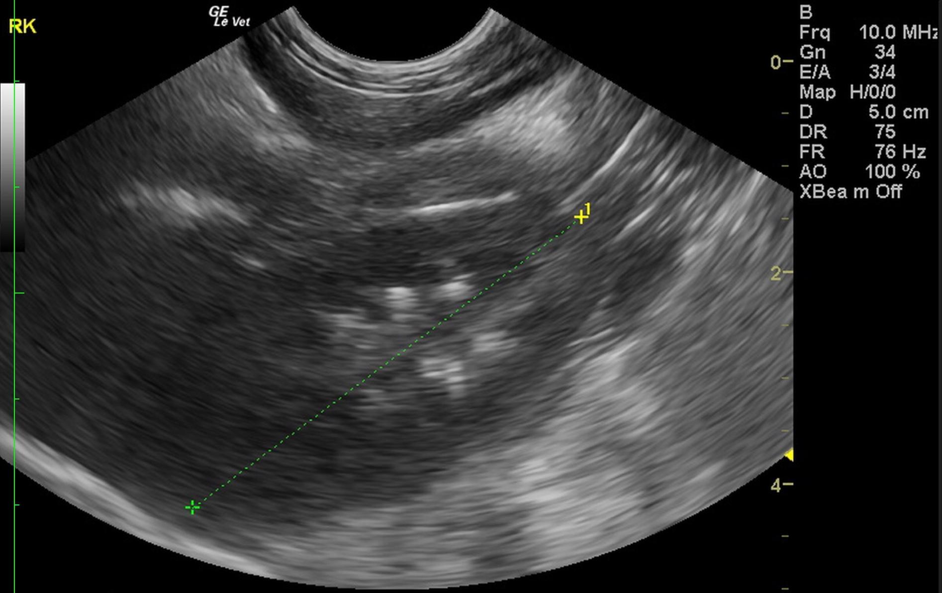
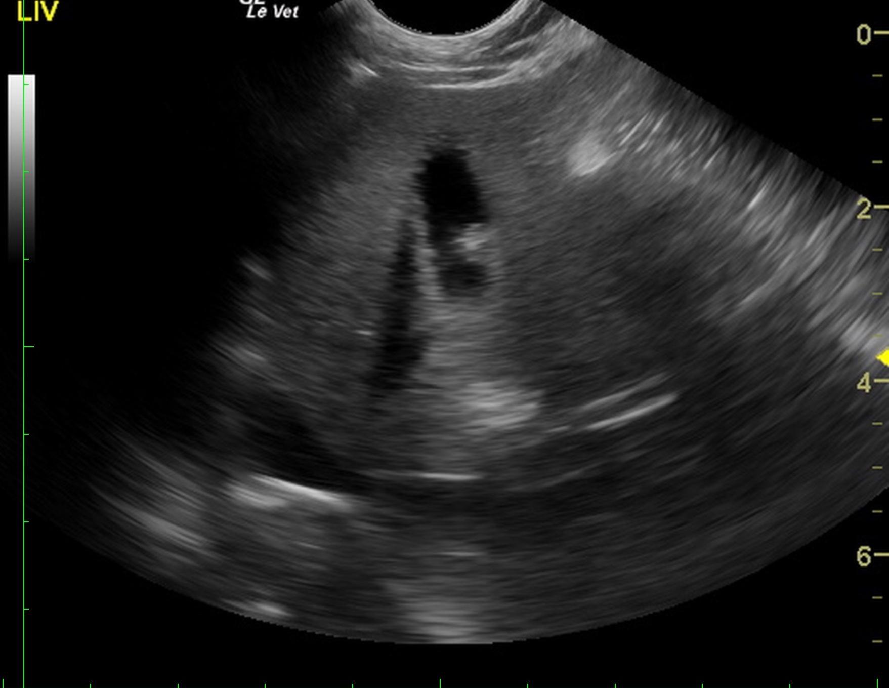
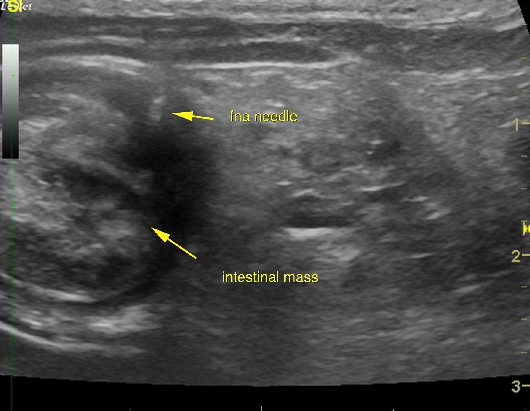
A 12-year-old, MN, Jack Russell terrier was presented for vomiting and weight loss. Radiographs revealed an abnormal gas pattern.
A 12-year-old, MN, Jack Russell terrier was presented for vomiting and weight loss. Radiographs revealed an abnormal gas pattern.
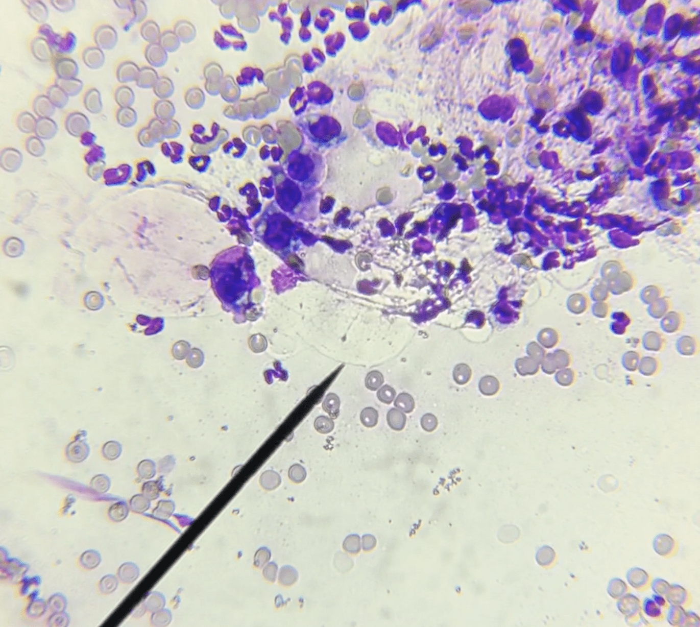
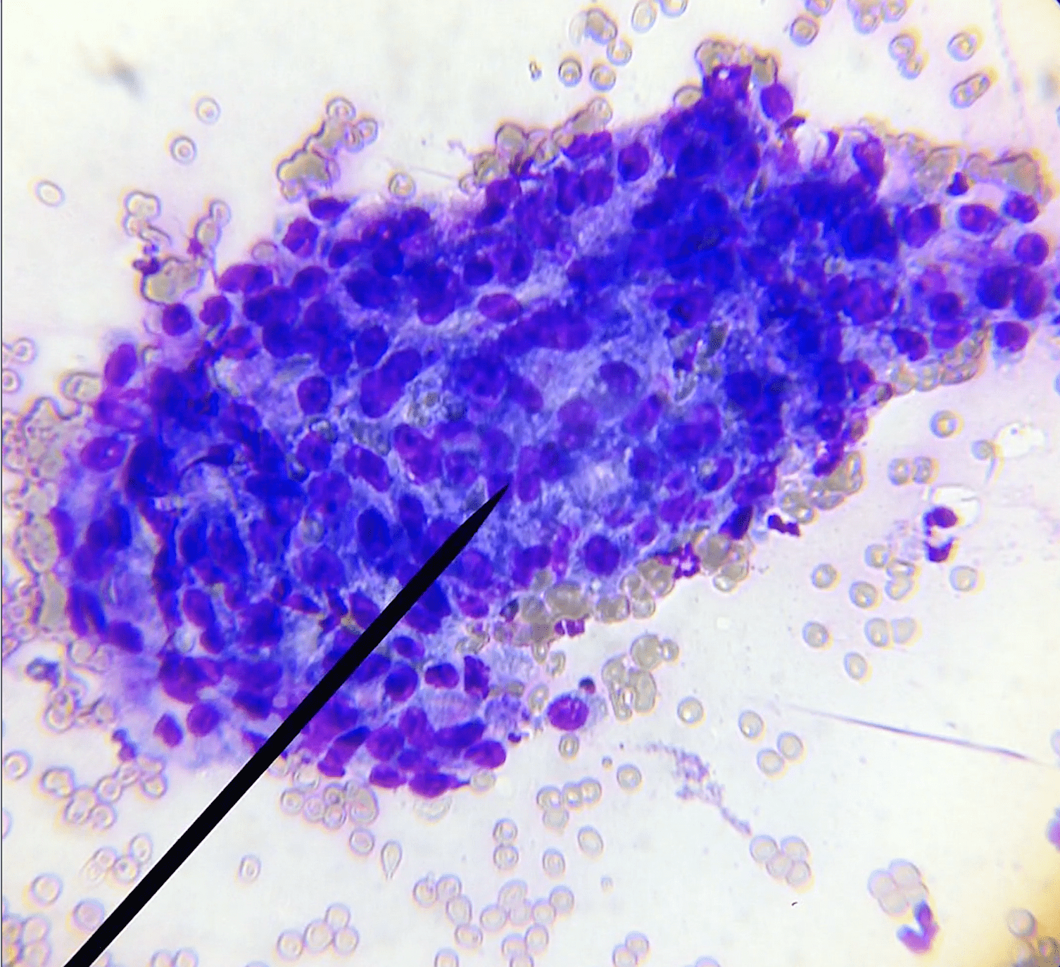
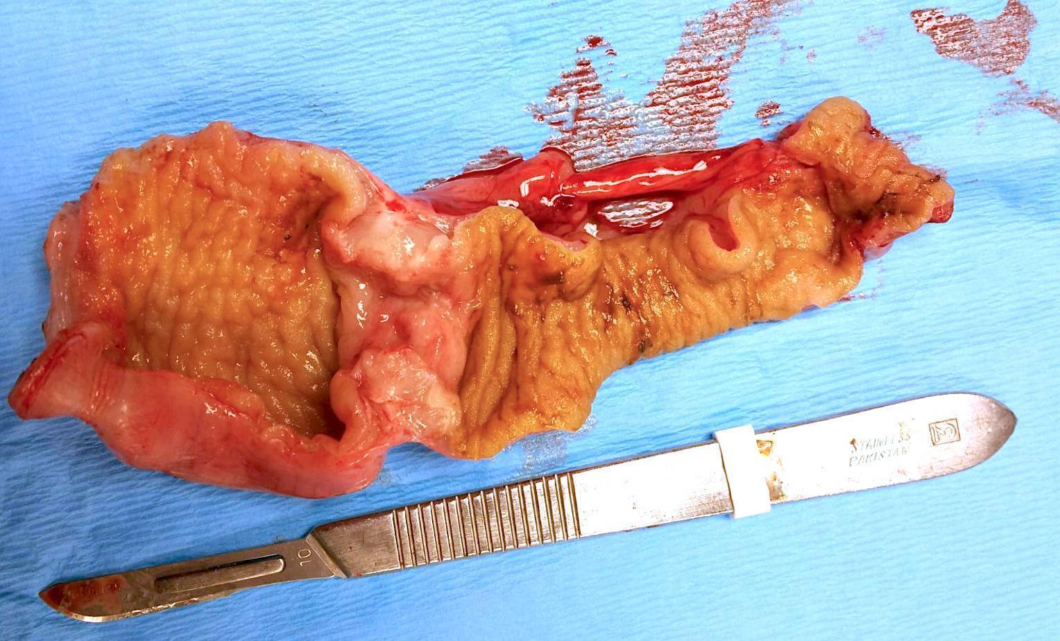
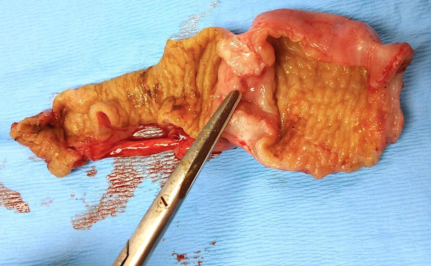
Intestinal mucoid carcinoma.
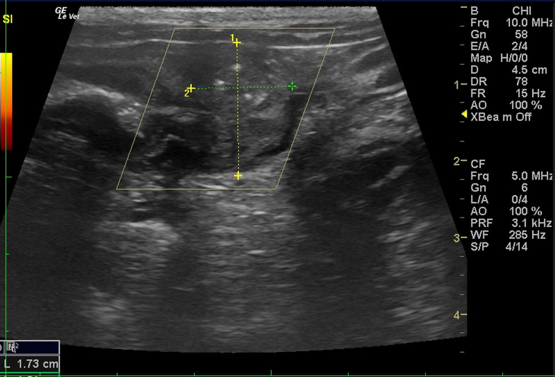
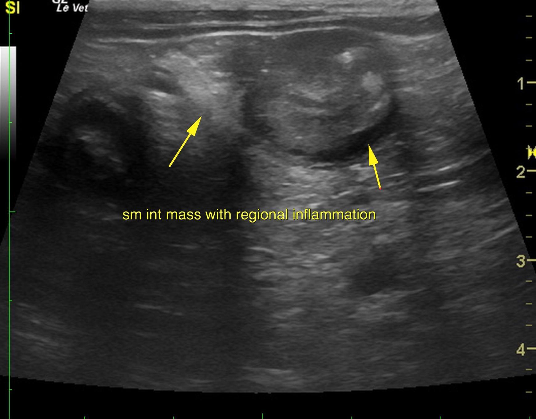
An annular, small intestinal mass was noted in the patient with focal areas of mineralization suggestive for intestinal carcinoma. This appeared partly obstructive. The intestinal mass appeared to be in the mid-small intestine/jejunum region. The mass measured approximately 1.5 to 2 cm.
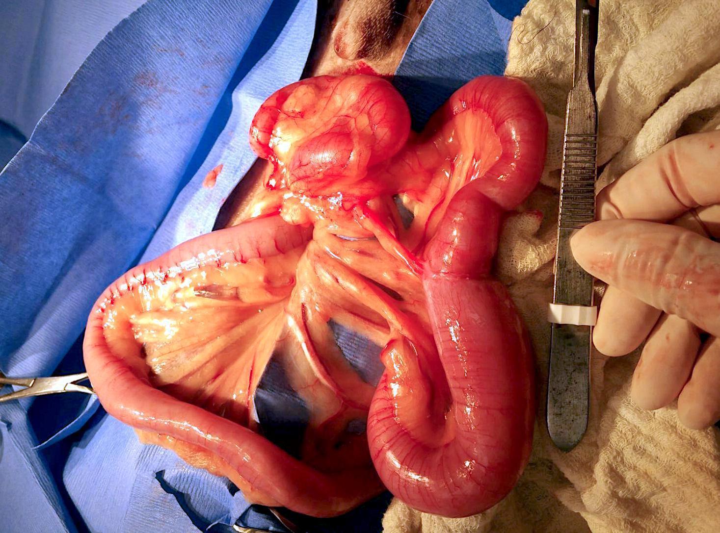
Cytology results: Inspissated mucus and supperative inflammation with a few irregular cells suggestive of intestinal mucoid carcinoma. Recommendations were for aggressive resection and anastamosis of the intestinal mass. The mass measured approximately 1.5 to 2 cm; however 6-8 cm of the intestine should be resected and anastamosed. 3-view chest radiographs were advised to assess for metastasis. The patient underwent surgery with resection and anastamosis and recovered from the procedure without event. At last follow up, the patient was on chemotherapy (tyrosine kinase inhibitor/piroxicam), doing very well with a normal appetite.