Being a young, very sweet and rambunctious dog with acute abdominal pain doesn’t always add up to a foreign body. Sometimes it can be a sign of something more ominous and sometimes it’s not what you thought at all. A positive Murphy sign during ultrasound with suspicion of possible foreign body vs. mass vs. other were the clinical differentials for this Case Of the Month. Many thanks to Dr. Edie Demaria of Pets Aloud Veterinary Services and her wonderful team in managing this case. SonoPath Mobile’s Kelly Vazquez, C.V.T., SDEP Certified Clinical Sonographer provided the diagnostic images for this case study.
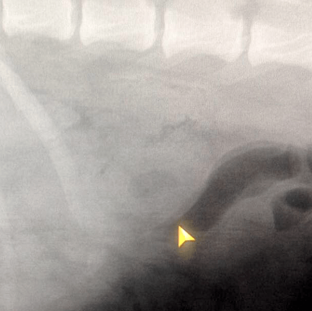
A 3-year-old, 60 lb, MN, Labrador Retriever mixed breed canine was presented with a history of possible dietary indiscretion while on a hike with his owner 48 hrs prior. The patient was ADR and had been intermittently vomiting for 24 hrs. Upon physical examination the patient exhibited pain in the caudal abdomen and an abnormal structure could be felt on palpation. Radiographs showed an irregular, semi-circular area in the caudal abdomen. The patient was hospitalized on I.V. fluids and supportive care pending ultrasound.
A 3-year-old, 60 lb, MN, Labrador Retriever mixed breed canine was presented with a history of possible dietary indiscretion while on a hike with his owner 48 hrs prior. The patient was ADR and had been intermittently vomiting for 24 hrs. Upon physical examination the patient exhibited pain in the caudal abdomen and an abnormal structure could be felt on palpation. Radiographs showed an irregular, semi-circular area in the caudal abdomen. The patient was hospitalized on I.V. fluids and supportive care pending ultrasound.
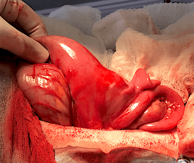
Intramural abscess.
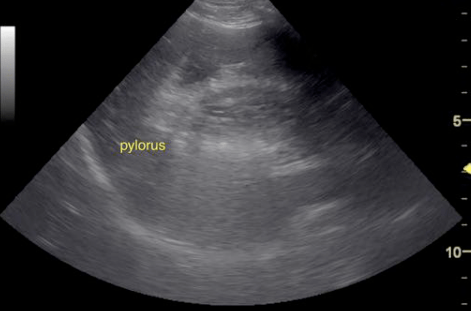
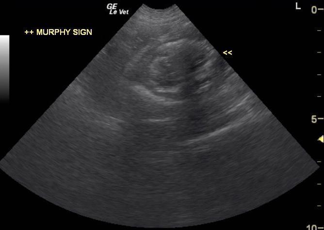
A portion of bowel in this patient appeared to be thickened and possibly intussuscepted measuring 4.0 cm with variable mural thickening that measured approximately 1.0 cm. The entire structure measured approximately 4-5 cm with shadowing material. The structure is likely fluid absorbing or passage of fluid is occurring as there is not a large, obstructive fluid pattern prior to the pathology. Intestinal mass or intussusception is suspected with embedded luminal material.
The area of concern for intestinal mass, intussusception, or possible foreign body appeared to be jejunum, yet the architecture was lost in portions of the structure and therefore the exact location of the pathology was unclear. There was a portion, approximately 1.5 cm, in the lumen that would be consistent with foreign matter or possible tissue proliferation. However, it did appear luminal so there was a possibility for the presence of foreign matter with secondary intussusception. Exploratory surgery was recommended with the likelihood of resection and anastamosis of the intestines. At the time of surgery an intramural abscess was discovered at the ileocolonic junction. Biopsy results were the following: Diverticulum with rupture and multifocal mural pyogranulomatous necro-hemorrhagic proliferative enterocolitis and peritonitis. No overt evidence of neoplasia was seen. Culture from the area found heavy growth of E. Coli. The patient recovered well post-op and was doing wonderfully at his suture removal follow-up exam.