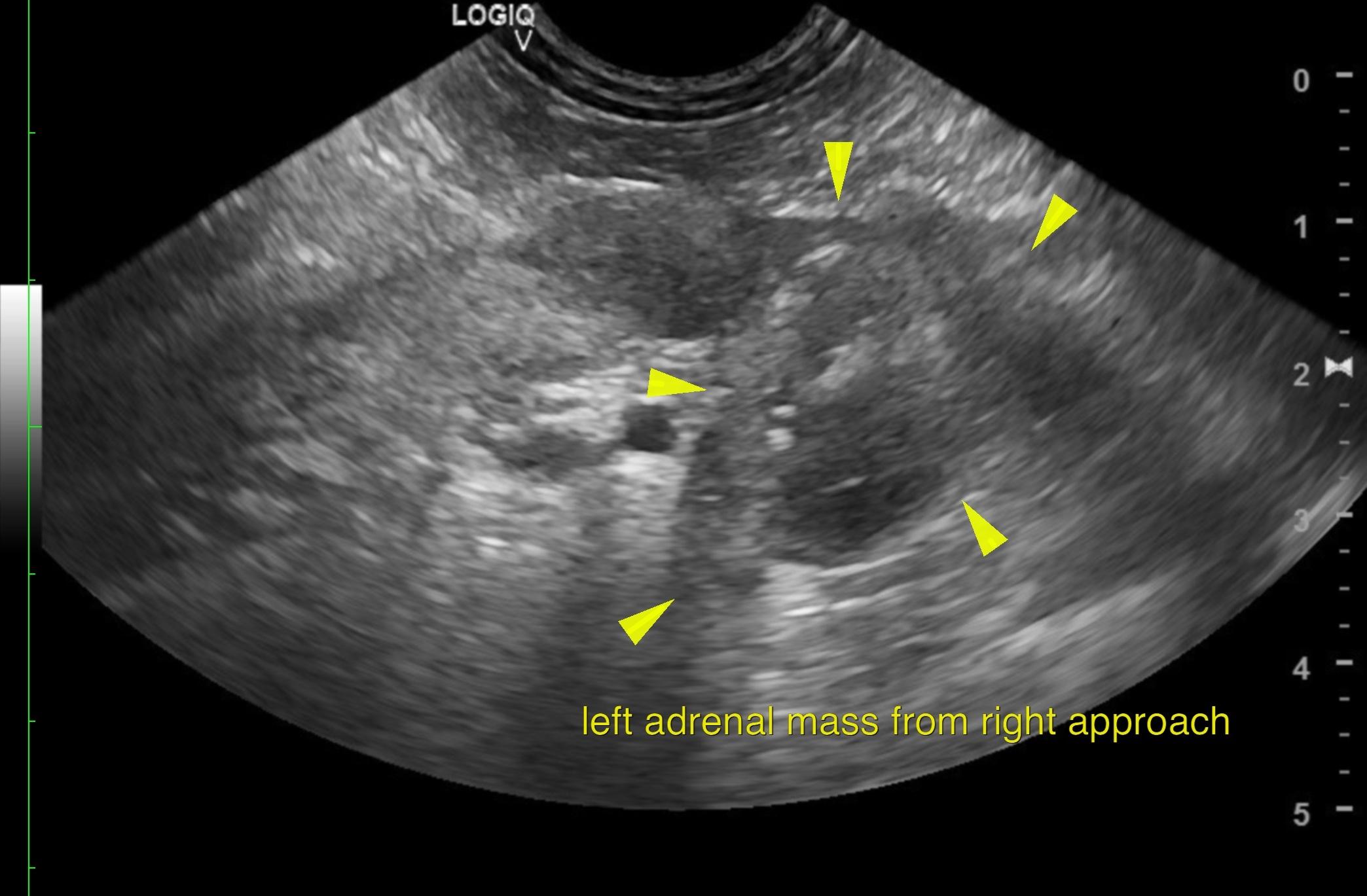
Veterinary technicians and assistants can be very competent sonographers, as we see here on this impressive diagnostic imaging by veterinary assistant Carly Pate. Carly’s modification of position 14 of the SDEP 17 point abdominal progression enabled imaging of the left adrenal from the right side, thus acquiring this definitive diagnostic image of CVC invasion of the left adrenal mass. Non-specific clinical signs always warrant a complete abdominal scan, and now with a specific treatment direction this dog is off to surgery. Many thanks to Dr. Ester Kastella for the management of this challenging case, and to the staff of VCA McKenzie Animal Hospital for their continued care of this patient.
The patient originally presented with a swelling of the lower right palpebrae, a 2 week history of hyporexia, change in treat preference, lethargy, decreased water intake and 2-3lbs of weight loss. Patient began drooling soon after he was sent home on Cefpodoxime, Apoquel and Mycequin for ocular changes. Patient has also been receiving NeoPolyBac ointment along the eyelid.
The patient originally presented with a swelling of the lower right palpebrae, a 2 week history of hyporexia, change in treat preference, lethargy, decreased water intake and 2-3lbs of weight loss. Patient began drooling soon after he was sent home on Cefpodoxime, Apoquel and Mycequin for ocular changes. Patient has also been receiving NeoPolyBac ointment along the eyelid.
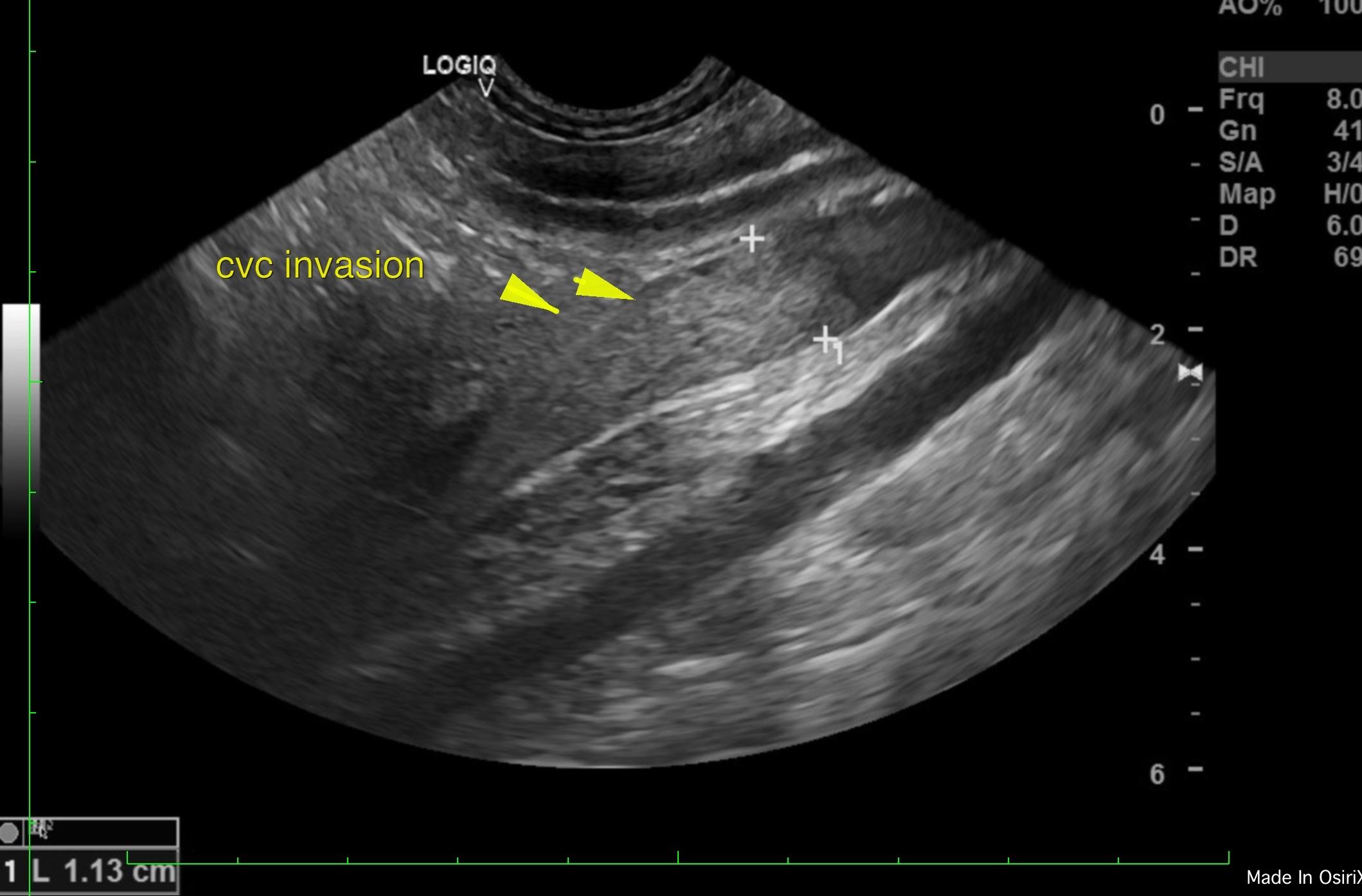
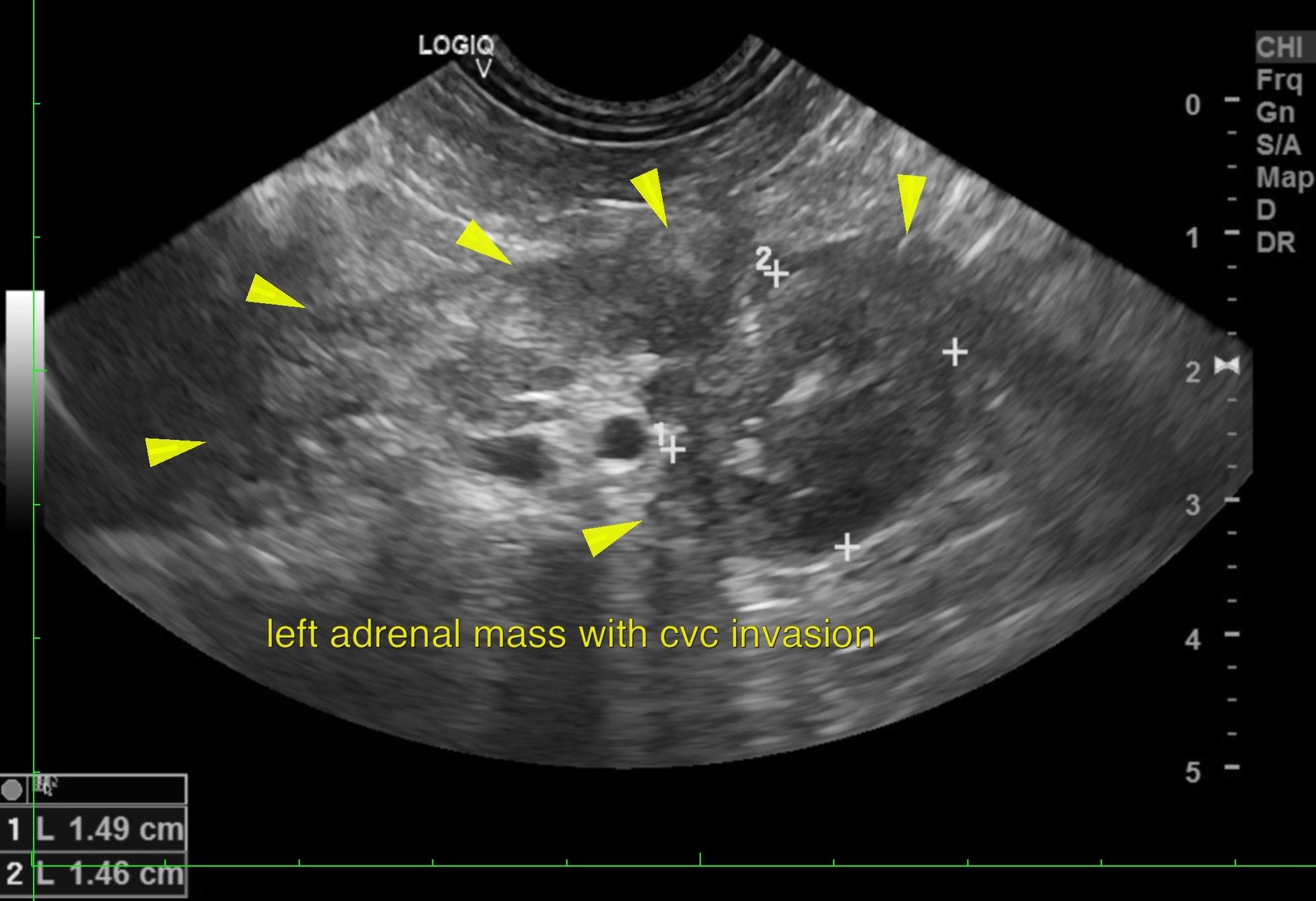
Mineralized left adrenal gland mass with attached thrombus/invasion into the vena cava.
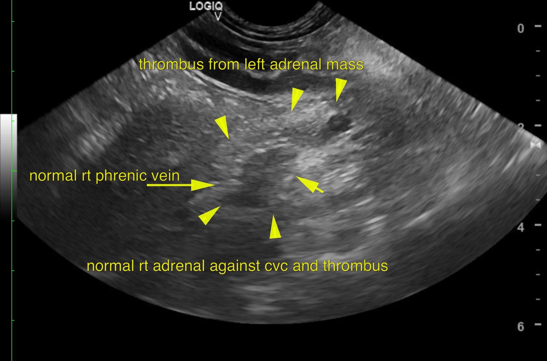
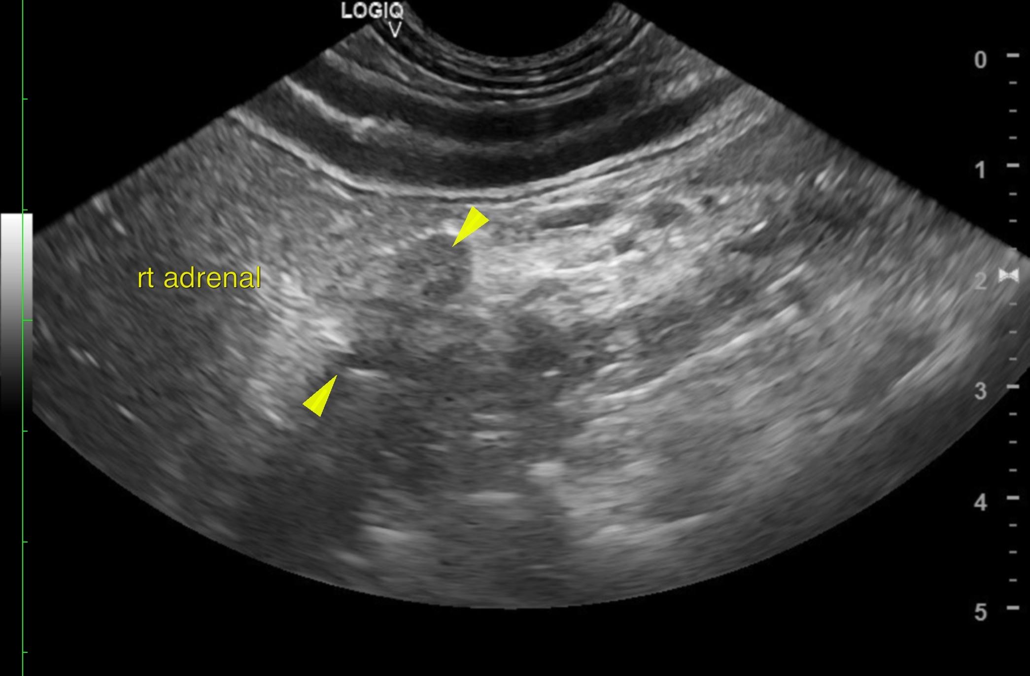
Left adrenal gland masses measuring 1.72 cm x 1.57 cm were noted, and seen invading the vena cava when visualized from the right approach; the vena cava was invaded for approximately 4 cm. The mineralizations seen would suggest carcinoma. The right adrenal gland was recognized as having normal shape, size, position and echogenicity for this breed. The phrenic vasculature, glandular echogenicity and detail were unremarkable. Capsule, cortex, and medullary definition were normal for this age patient. The right adrenal gland measured 1.5 cm in length and 0.5 cm in width.
A surgical approach is a potential, if the surgeon is prepared for thrombus-occupation of the vena cava to an extent of approximately 4 cm cranial to the left adrenal mass. No overt evidence of metastatic disease was noted. Surgical consultation was recommended. The mineralization suggested carcinoma. The patient was sent to a referral facility for surgical consultation and was scheduled for left adrenalectomy with removal of the associated vena cava thrombus.