A 13-year-old MN Beagle mix with history of increased weakness and respiratory effort presented for a heart murmur, suspected pulmonary edema, enlarged liver, and possible cranial abdominal mass. The patient was abdominally retracting while breathing, but his mucous membranes remained consistently pink. The patient was started on furosemide 12.5 mg 3 tabs BID and Pimobendan 5 mg BID. CBC and blood chemistry found moderately high WBC count; poss. bands, Alk. Phos. 663. Urine specific gravity was 1.023.
A 13-year-old MN Beagle mix with history of increased weakness and respiratory effort presented for a heart murmur, suspected pulmonary edema, enlarged liver, and possible cranial abdominal mass. The patient was abdominally retracting while breathing, but his mucous membranes remained consistently pink. The patient was started on furosemide 12.5 mg 3 tabs BID and Pimobendan 5 mg BID. CBC and blood chemistry found moderately high WBC count; poss. bands, Alk. Phos. 663. Urine specific gravity was 1.023.
Myocardial sarcoma pattern involving left ventricle, left atrial free wall, and right ventricle. Probable concurrent splenic involvement.
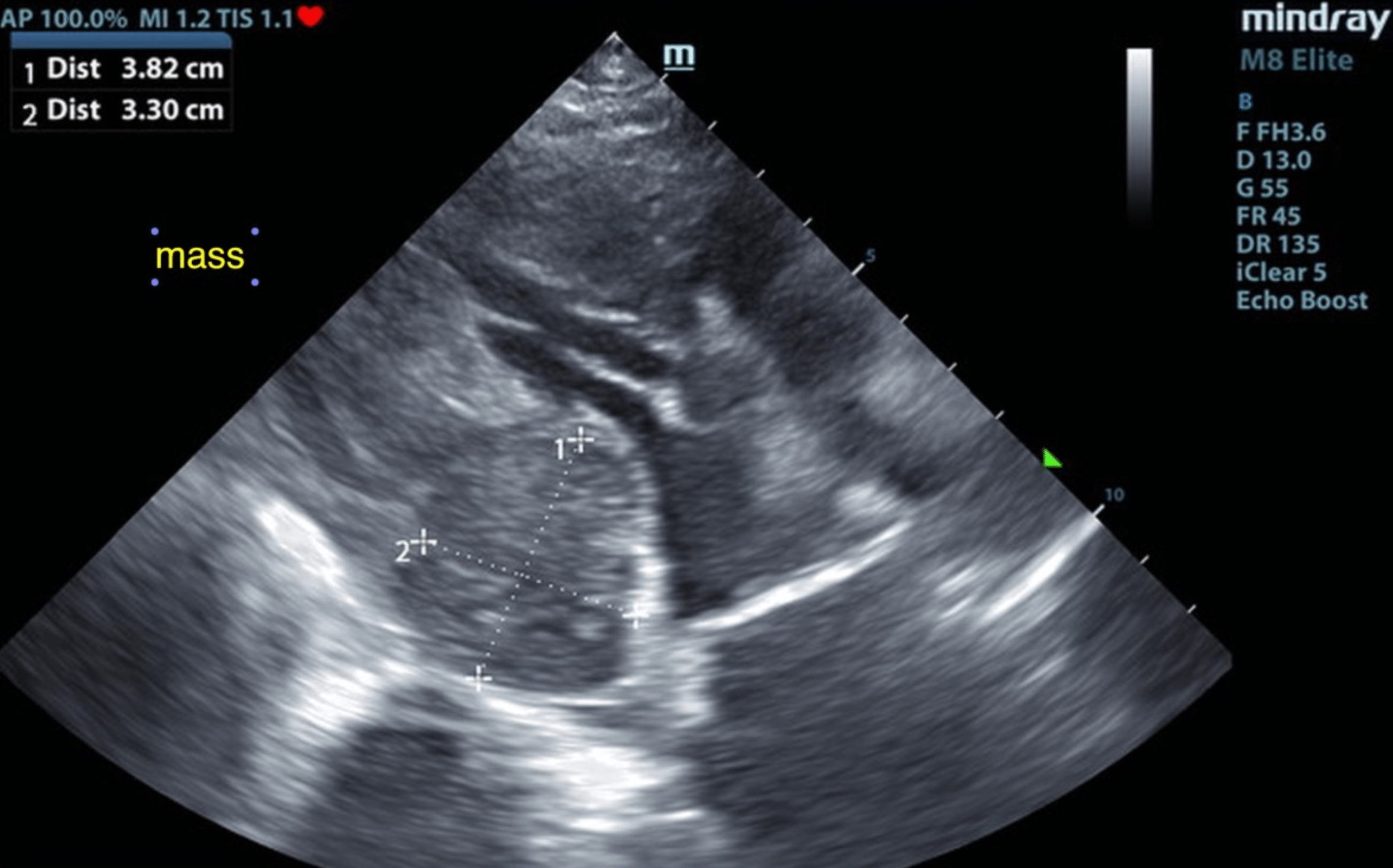
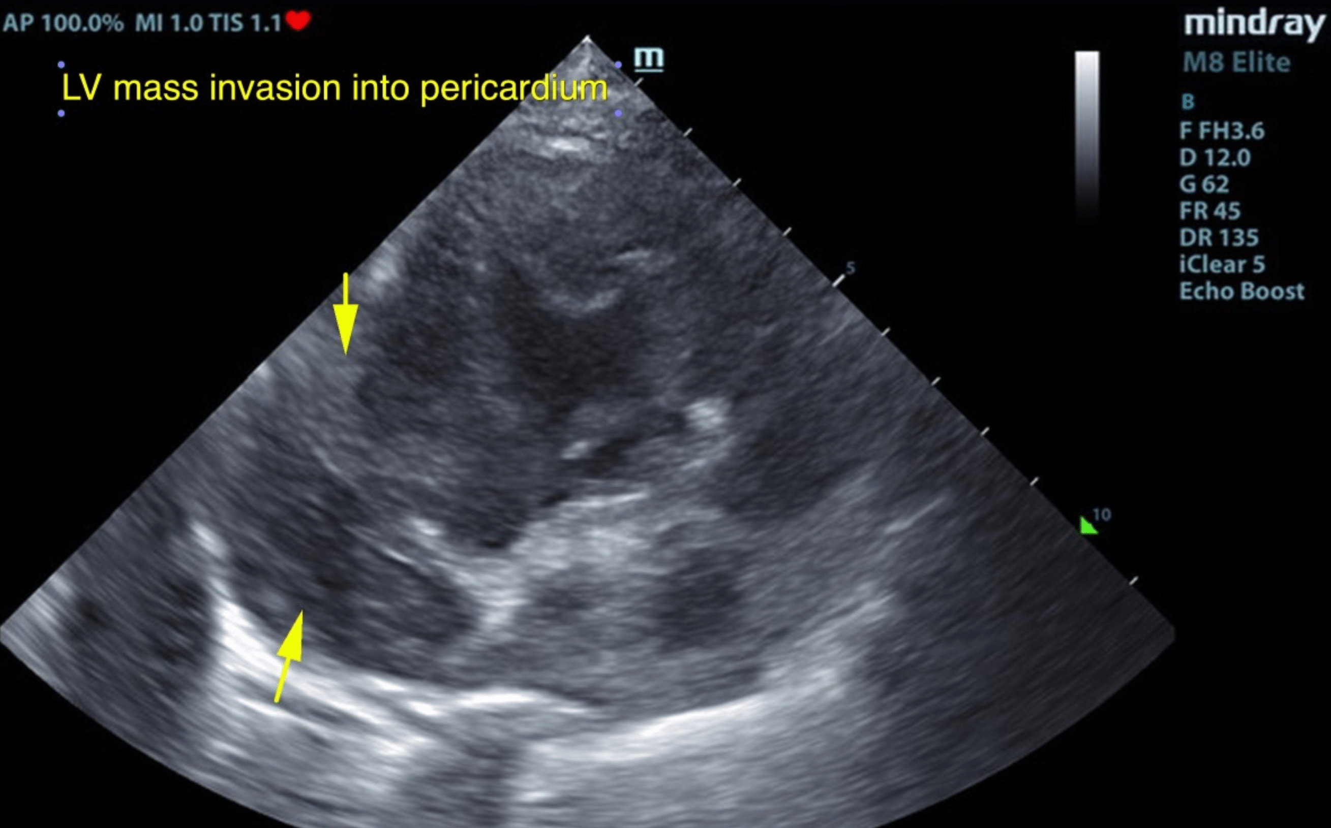
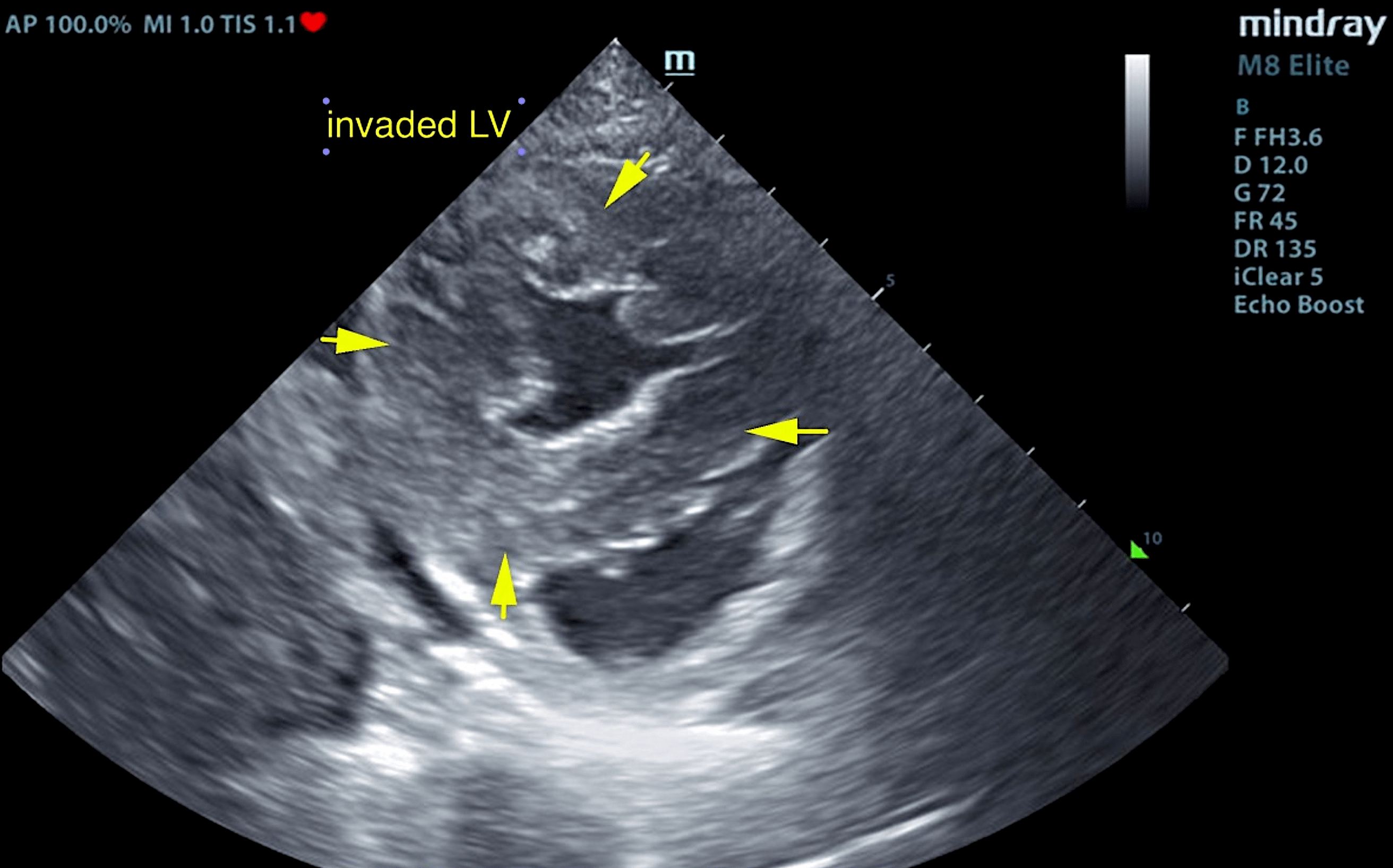
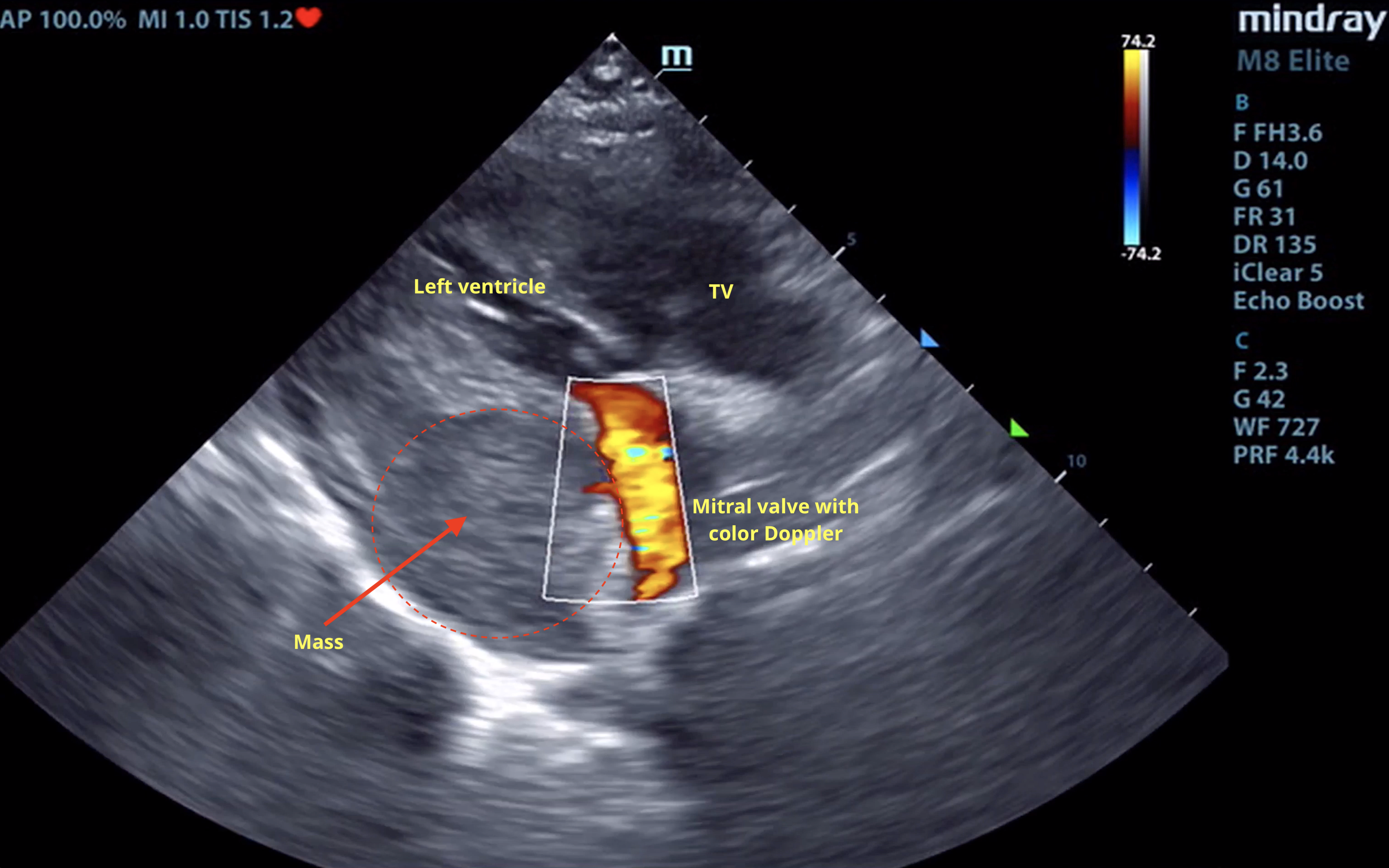
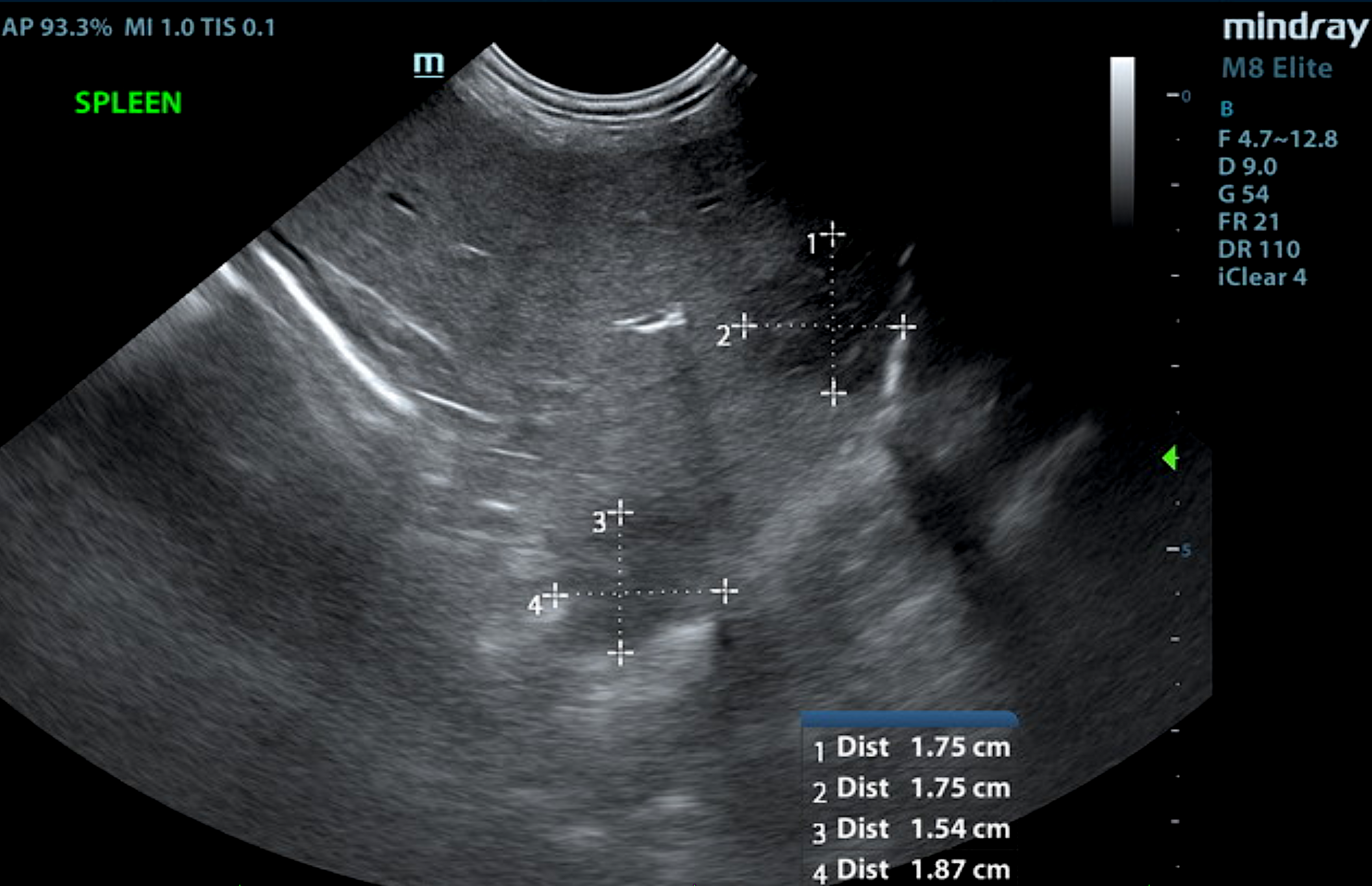
The left ventricle septum and left ventricular free wall in this patient revealed infiltrative, mixed hypoechoic tissue proliferation and culminating in a focal 3.8 cm mass at the junction of the left ventricular free wall and the left atrium. This pattern is strongly consistent with myocardial sarcoma. Impingement of the mass into the left atrial lumen was noted. Some infiltrative pattern also continued into the right ventricle. Myocardial sarcoma pattern involving left ventricle, left atrial free wall, and right ventricle. The mass extends into the pericardium. A slight amount of pericardial effusion was noted. Concurrently the spleen revealed multi-focal, hypoechoic nodules measuring up to 1.87 cm with disrupted architecture and target type appearance, likely related to the myocardial pathology.
FNA of the splenic nodules was recommended and immediate chemotherapeutic intervention with followup
echocardiogram. The prognosis is poor long term. Unfortunately, this patient was too compromised to survive long enough for any kind of treatment and passed away a few short days after diagnosis.