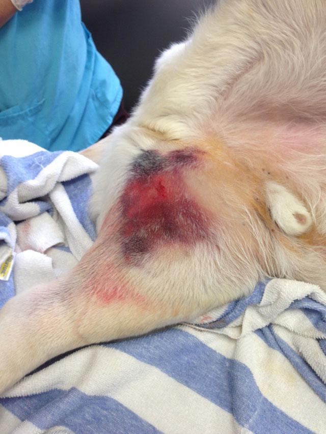
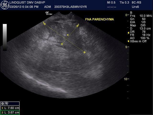
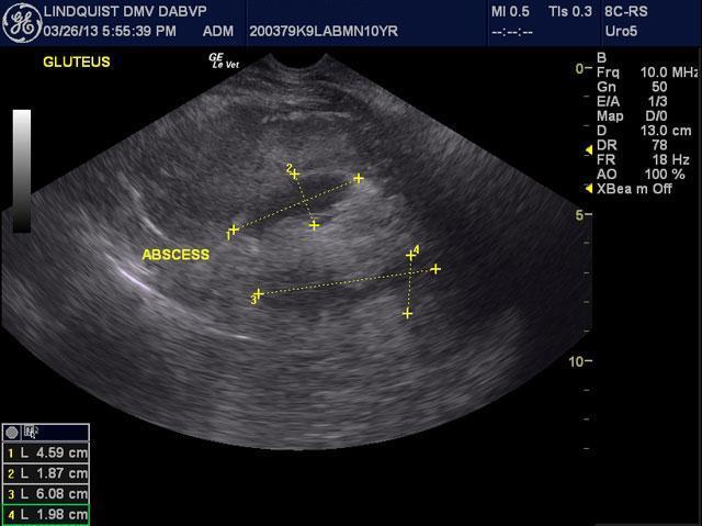
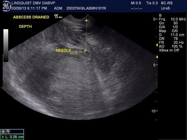
Sonograms aren’t just for the belly and chest. If we don’t know what it is, we put a probe on it, sample it, treat it, and put it on a slide. See this poor Labrador with an impressive swelling in the hind limb of unknown origin in the May, 2013 SonoPath case of the month. That’s why we have a probe, a needle, and diagnostic efficiency at SonoPath.com.
Sonogram (Pancreas): Gunner (Name changed to protect the innocent 🙂
History: A 7-year-old MN Labrador Retriever was presented for severe hind limb swelling, bruising in the inguinal region and grade 3 limp. Mild fever was present. Radiographs revealed severe soft tissue swelling of around the femur without bone involvement. Thoracic and abdominal radiographs were unremarkable.