Pyometra with male genitalia? An 8-month-old intact male Basset hound was scheduled for a routine neuter procedure but prior to the procedure, he presented for painfully swollen testicles. Externally the patient had normal male genitalia with the exception of large, swollen testicles. Internally the patient had fluid filled tubular structures resembling what appeared to be a pyometra, a surprising thing to see in male dog. Images for this case were provided by Kelly Vazquez, CVT, SDEP® certified clinical sonographer for SonoPath Mobile Veterinary Ultrasound with interpretation by Dr. Eric Lindquist, Founder & CEO of SonoPath.com, Director of SonoPath Mobile, NJ, Animal Sounds NW Mobile, Eugene Oregon, and Director of The Focal Zone, Ontario, Canada. Excellent care and patient management by Dr. Jill Shiffman, (owner of Bergen County Veterinary Center, Waldwick, NJ) and her compassionate team.
The patient presented for ADR, not eating well, progressive lethargy, and swollen testicles. The owner noted that his testicles had always been large and that he does have some degree of dietary indiscretion. The patient received buprenorphine and SQ fluids pending ultrasound interpretation.
The patient presented for ADR, not eating well, progressive lethargy, and swollen testicles. The owner noted that his testicles had always been large and that he does have some degree of dietary indiscretion. The patient received buprenorphine and SQ fluids pending ultrasound interpretation.
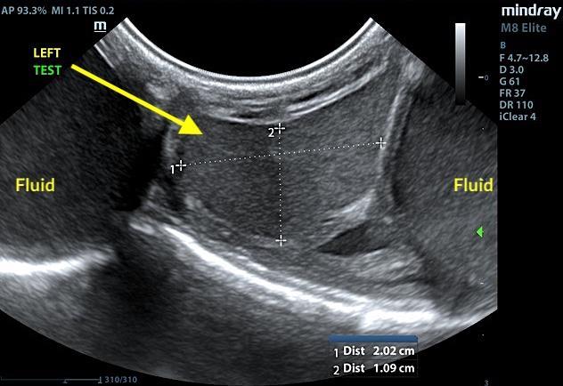
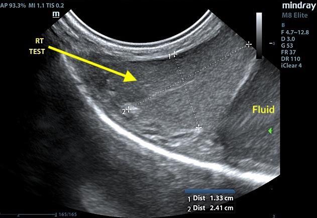
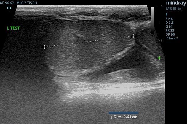
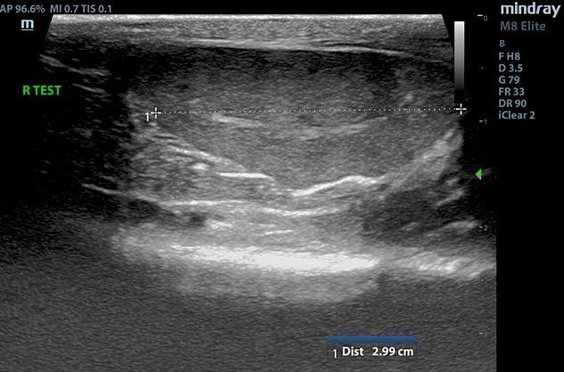
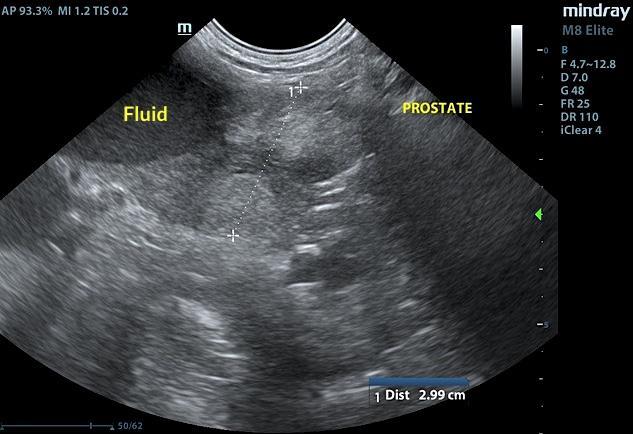
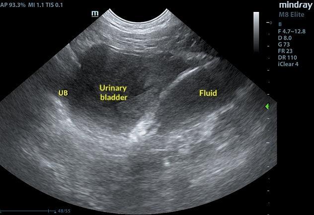
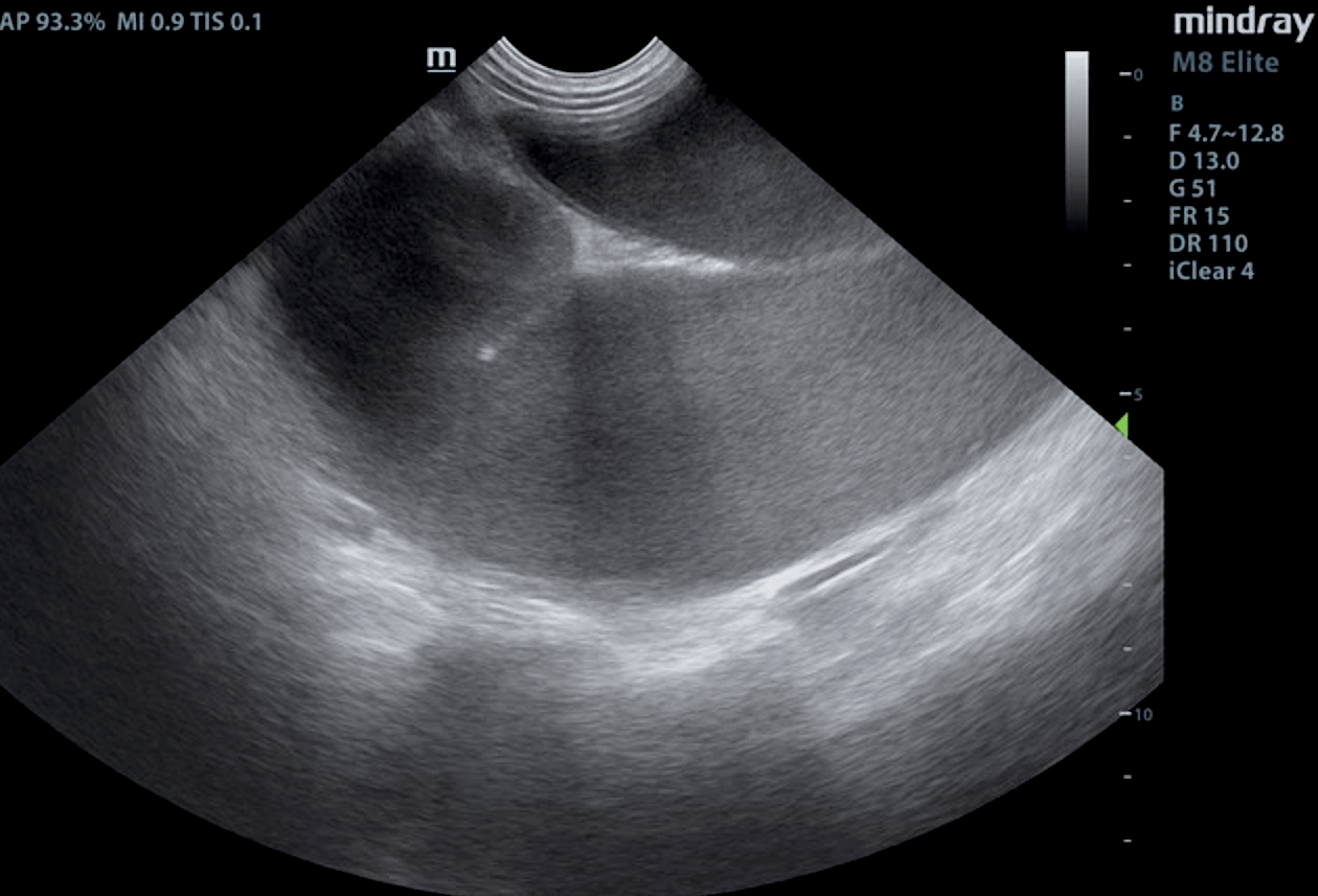
Prostatitis pattern with caudal abdominal tubular structures, possible pyometra owing to hermaphroditism.Fluid filled scrotal sac with structurally normal testicles.
The left testicle was uniform and measured 2.64 cm. The right testicle measured 2.4 cm and was uniform. The prostate was slightly enlarged and measured 3.0 cm with minor, heterogenous parenchymal changes. The prostate revealed striating edema lines. This is consistent with prostatitis. A large amount of free fluid was noted within the testicular sac. Echogenic fluid filled tubular structures were noted in the caudal abdomen. The material in the tubular structures appeared to be echogenic and likely purulent. These encompassed the urinary bladder and were in the region of the uterus. However, no ovaries were found. Hermaphroditism should be considered. Surgical exploratory would be warranted.
The patient was referred to an emergency surgical facility for immediate exploratory due to suspicion of persistent Müllerian duct syndrome vs. other. During surgery a fluid filled possible pseudo uterus was found; that structure was attached to either testes through a hernia in the body wall. A rare congenital condition known as persistent Müllerian duct syndrome was believed to be the finding. Patient is a pseudohemaphrodite. Both testicles were removed as was the fluid filled structure. Surgery notes: two inguinal hernias, communication from each of the testicles to a fluid filled structure in the abdomen, 800 mls of purulent filled was extracted from the fluid filled structure and neuter was performed. The patient is expected to recovered well after surgery and was treated with Unasyn, buprenorphine, and continued I.V. fluids post-op. The fluid filled structure and both testicles were submitted for biopsy and cultures to the lab.
Pathology results confirmed the presence of a tubular organ- oviduct and uterus.Pathology comments: the finding of a tubular organ that histologically resembles different areas of oviduct and uterus is compatible with the clinical diagnosis of a persistent Müllerian duct syndrome.Testicles: testis azoospermia with plexus malformation and moderate lymphoplasmacytic duct efferent ductitis.