Everyone wants to find a pheo… well here is a little 1 cm pheo 🙂 Isn’t she cute??…. but malignant. I always lecture that now that we are seeing adrenal glands every time in our SDEP ultrasound protocol it doesn’t mean that every adrenal nodule is a pheochromocytoma. Well, in this month’s case this little expansive nodule focally deviating the left adrenal capsule was, in fact, a pheo with the usual vague clinical signs on presentation. See how a pheo may present and how it may look in this month’s SonoPath case of the month imaged by Andi Parkinson, RDMS/Rachel Brilhart, RDMS of Intrapet Imaging, Baltimore, MD and interpreted by yours truly, Eric Lindquist, DMV, DABVP, Cert. IVUSS and Dr. Maggie Machen DVM, DACVIM (Cardiology) via SonoPath’s telemedicine services. Case managed by Dr. L. Zakai of Frederick Road Veterinary Hospital.
The patient was presented for heavy breathing, panting, excessive throat clearing, weight loss. Altered CBC/Chem/UA values: BW-NSF. Radiograph Findings: chest rads-NSF.
The patient was presented for heavy breathing, panting, excessive throat clearing, weight loss. Altered CBC/Chem/UA values: BW-NSF. Radiograph Findings: chest rads-NSF.
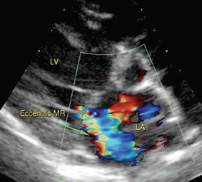
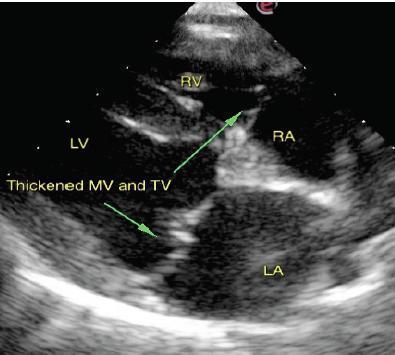
Abdominal ultrasound revealed a left adrenal gland nodule. Otherwise, unremarkable abdomen. Echocardiogram: Chronic degenerative valve disease causing moderate mitral and tricuspid regurgitation.
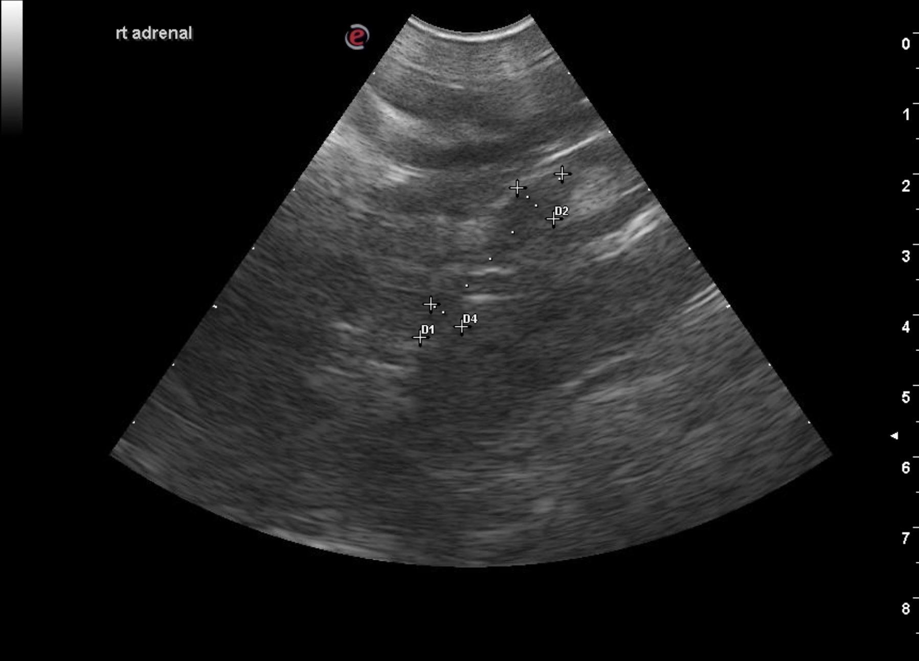
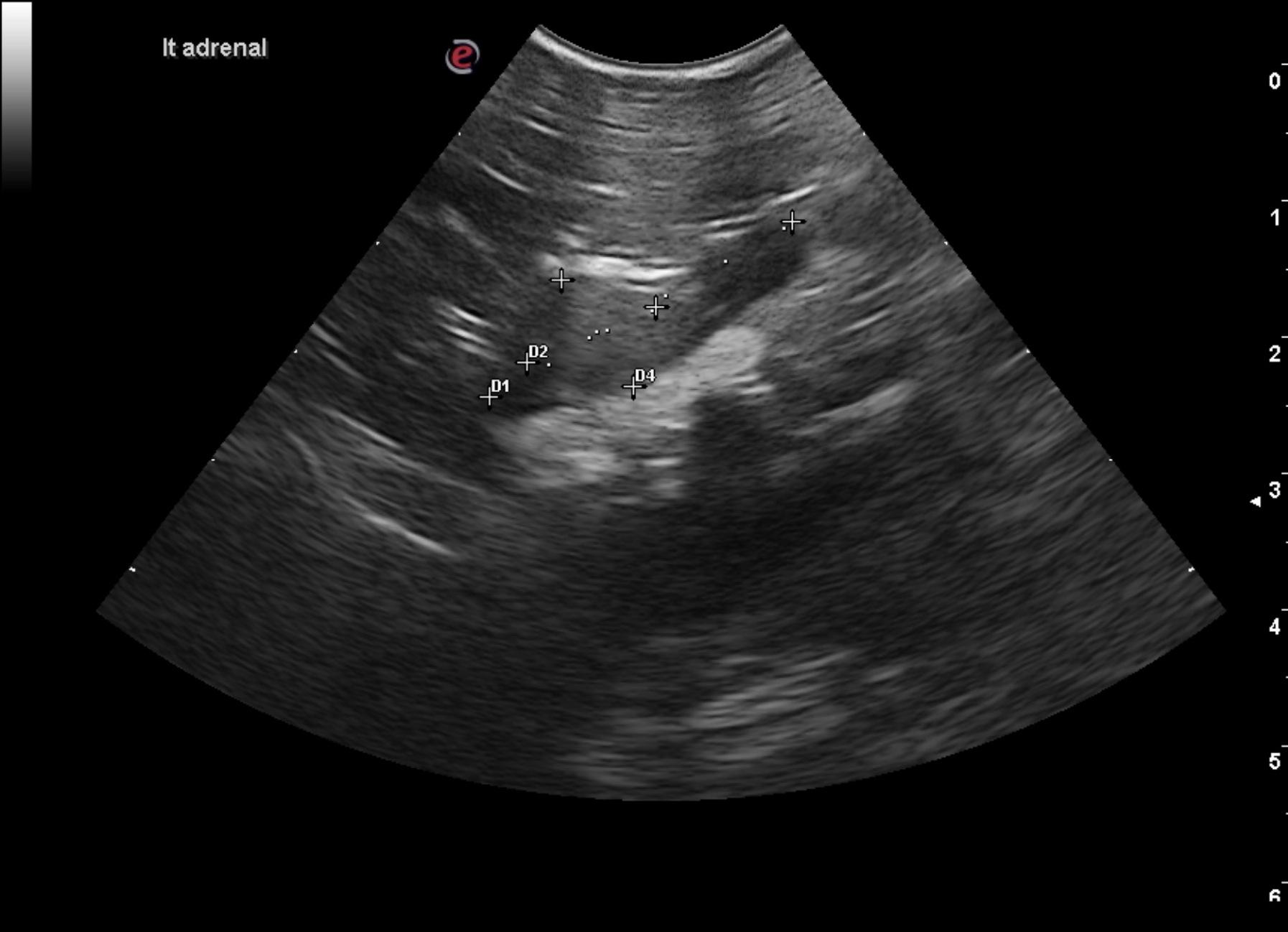
Abdomen: The left adrenal gland presented a hyperechoic 1.03 cm nodule at the mid cranial pole. The left adrenal gland measured 2.57 cm in length. The left adrenal gland nodule presented capsular expansion without capsular escape with no evidence of vascular invasion. This appears resectable. The right adrenal gland was uniform and measured 3.07 x 0.68 cm at the caudal pole and 0.54 cm at the cranial pole.
Echocardiogram: Chronic degenerative valve disease causing moderate mitral and tricuspid regurgitation.
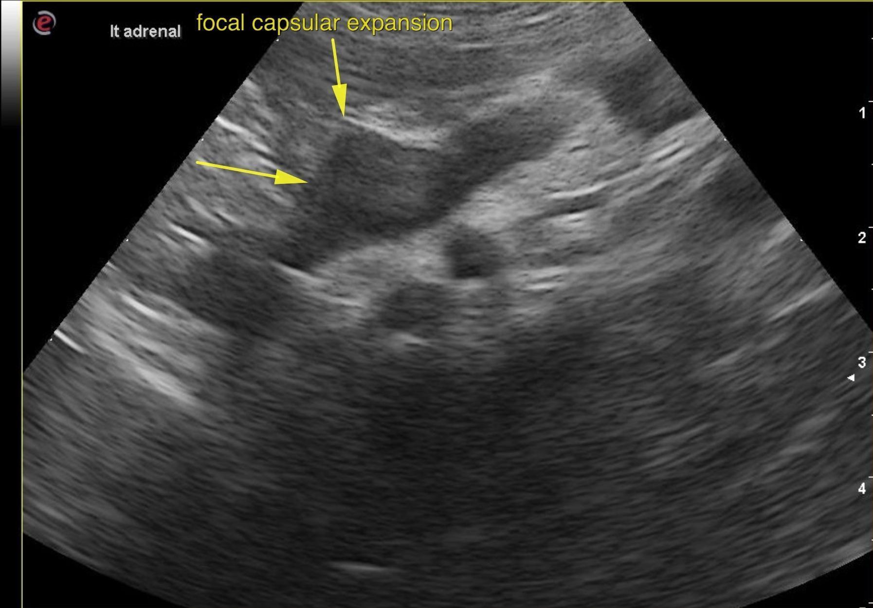
Serial blood pressure measurements +/- full adrenal panel was recommended. A recheck sonogram was recommended in 1-2 weeks to assess for any progression of the lesion. There is one portion of the adrenal gland nodule that is concerning and has focal, capsular expansion. Pheochromocytoma, adenocarcinoma and adenoma are all possible. Surgery found pheochromocytoma.