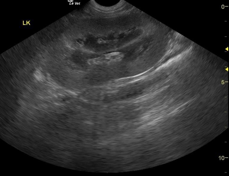
A 3-year-old male Labrador Retriever dog was presented for anorexia and weight loss. CBC was within normal limits, however blood chemistry showed hyperproteinemia, hypoalbuminemia, low albumin/globulin ratio, marked azotemia, hyperphosphatemia, mild hypocalcemia, hyperkalemia, and hyperamylasemia. T4 was within normal range. The urine had a cloudy appearance; 3+ proteinuria and 3+ hematuria were present on urinalysis.
A 3-year-old male Labrador Retriever dog was presented for anorexia and weight loss. CBC was within normal limits, however blood chemistry showed hyperproteinemia, hypoalbuminemia, low albumin/globulin ratio, marked azotemia, hyperphosphatemia, mild hypocalcemia, hyperkalemia, and hyperamylasemia. T4 was within normal range. The urine had a cloudy appearance; 3+ proteinuria and 3+ hematuria were present on urinalysis.