Whats are the 3 “Bears in the Forest” maneuvers on SDEP clinical sonography protocol?
Fresh off the SDEP abdomen course in Atlanta just two months ago, Dr. Karen Ebersole applied these SDEP maneuvers she learned over a weekend to this challenging intestinal mass hidden in the GI artifact. Give the patient the benefit of the doubt and don’t let the GI neoplastic criteria fool you; exploratory and histopath revealed that what looks like a duck and quacks like a duck may be something else. See what Dr Ebersole and her veterinary posse found in this dog with a GI mass obstruction.
Many thanks to Dr. Karen Ebersole of Lisbon Road Animal Hospital for the diagnostic ultrasound images and wonderful management of this case.
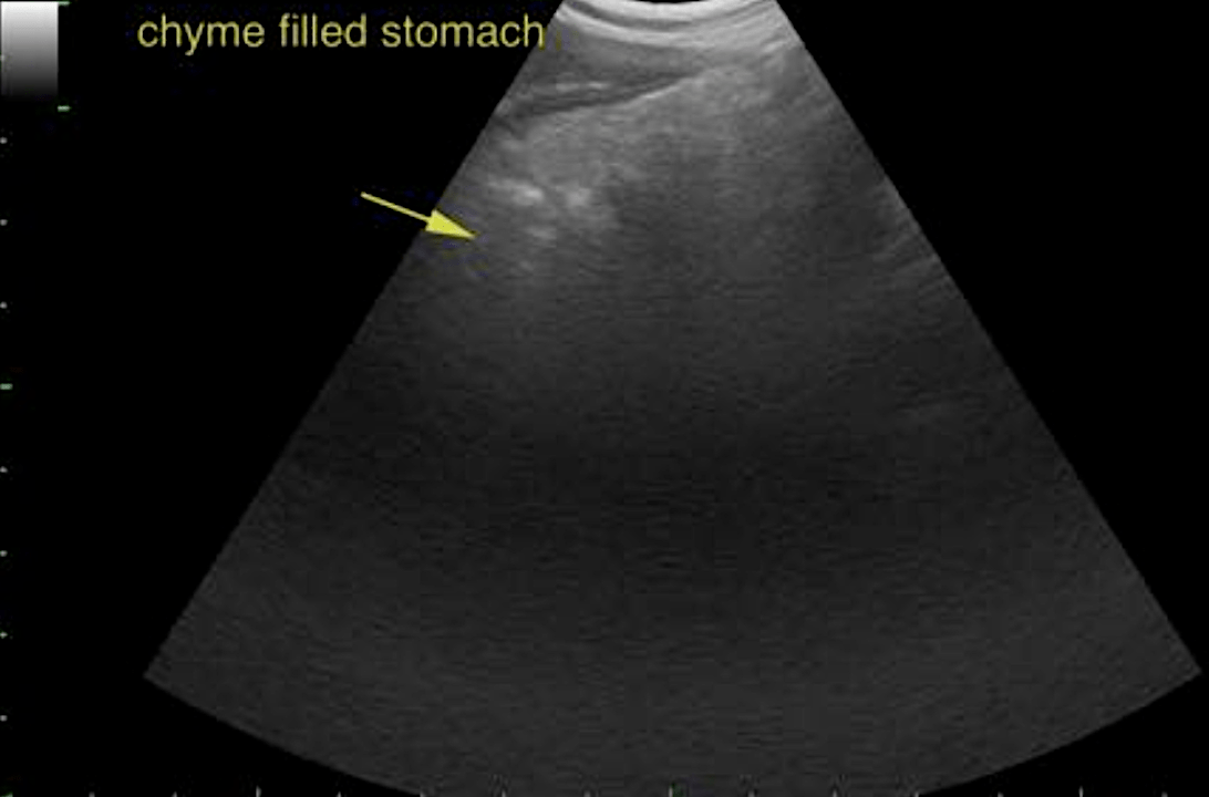
A 3-year-old, MN, Catahoula Leopard Dog was presented for a 4 month history of ADR, diarrhea, and vomiting which had acutely worsened within the last 48 hours. Physical examination found the patient with a body score of 2/9 with a 14 lb weight loss, temperature of 102.9, QAR, and mildly dehydrated. CBC/Chem, cPLI, and fecal were all WNL with the exception of an increase in monocytes (11%) with a normal WBC of 14,650. A full abdominal ultrasound was performed.
A 3-year-old, MN, Catahoula Leopard Dog was presented for a 4 month history of ADR, diarrhea, and vomiting which had acutely worsened within the last 48 hours. Physical examination found the patient with a body score of 2/9 with a 14 lb weight loss, temperature of 102.9, QAR, and mildly dehydrated. CBC/Chem, cPLI, and fecal were all WNL with the exception of an increase in monocytes (11%) with a normal WBC of 14,650. A full abdominal ultrasound was performed.
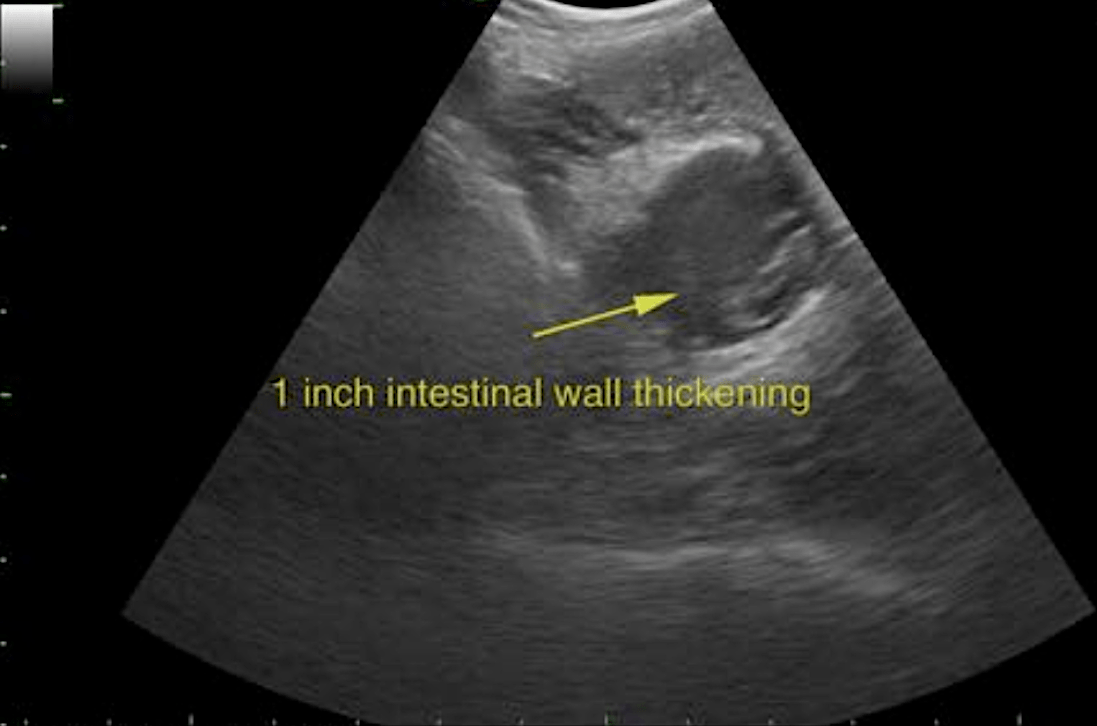
Jejunal mass: Severe, multifocal suppurative pyogranulomatous transmural enteritis. Mesenteric lymph node: Reactive.
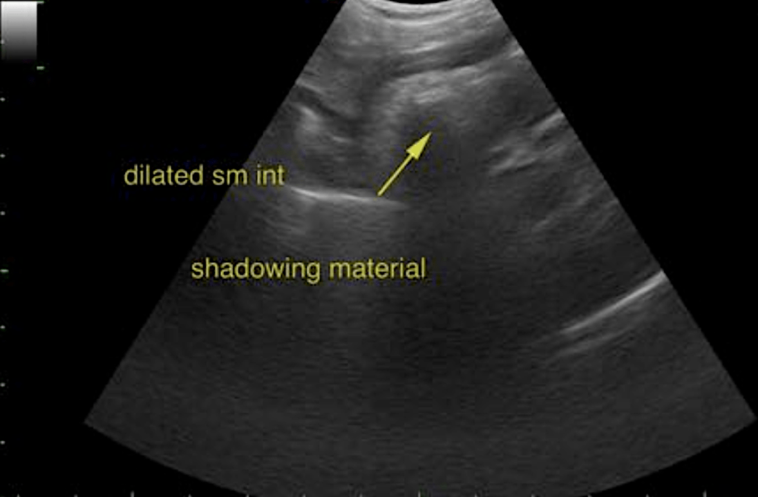
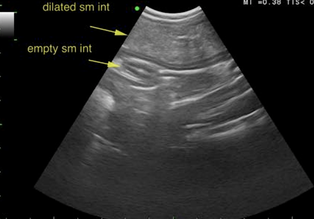
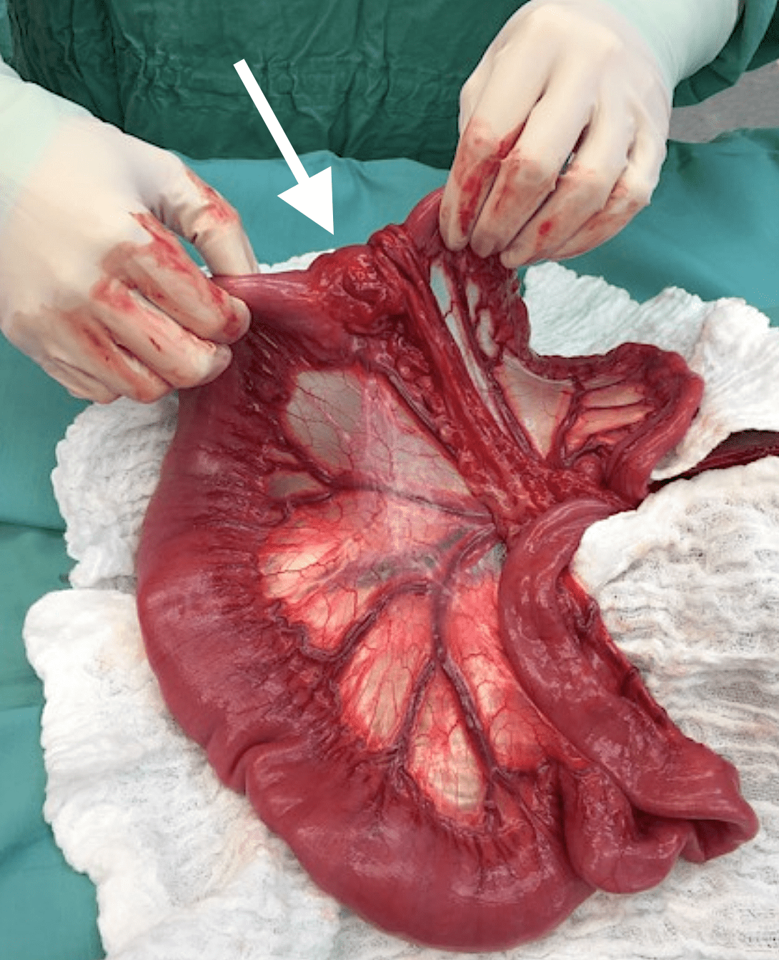
An abdominal exploratory was performed and a 1 inch jejunal mass was found causing an almost complete G.I. obstruction. A resection and anastamosis was performed with 18 inches of small intestine removed. The mass along with a section of mesenteric lymph node was sent to histopathology. The patient recovered very well post-operatively. He was treated with IV fluids and antibiotics, for the next 48 hours. At staple removal the patient was found to be in excellent recovery , with no vomiting or diarrhea, and several pounds of weight gain.