Traumatic event or emerging hemangiosarcoma? Note the diagnostic power of a recheck sonogram. Giving the benefit of the doubt is best for the patient but rechecking for regression or progression of the lesion will enhance your gut feeling for the underlying pathological truth. Check out this presentation imaged by Andi Parkison RDMS of Intrapet Imaging, Baltimore, Maryland, USA and how a prudent follow-up was key to the true diagnosis in this case despite the “traumatic’ history. A special thanks to Hickory Veterinary Hospital, Dr. Melissa Adams and Dr. Bob Silcox.
A 5-year-old male German shepherd was presented for evaluation of progressive pain, lethargy, anorexia, and hunched back following an episode of abdominal trauma – tried to jump over a large hole and hit his abdomen on the edge of the hole.
A 5-year-old male German shepherd was presented for evaluation of progressive pain, lethargy, anorexia, and hunched back following an episode of abdominal trauma – tried to jump over a large hole and hit his abdomen on the edge of the hole.
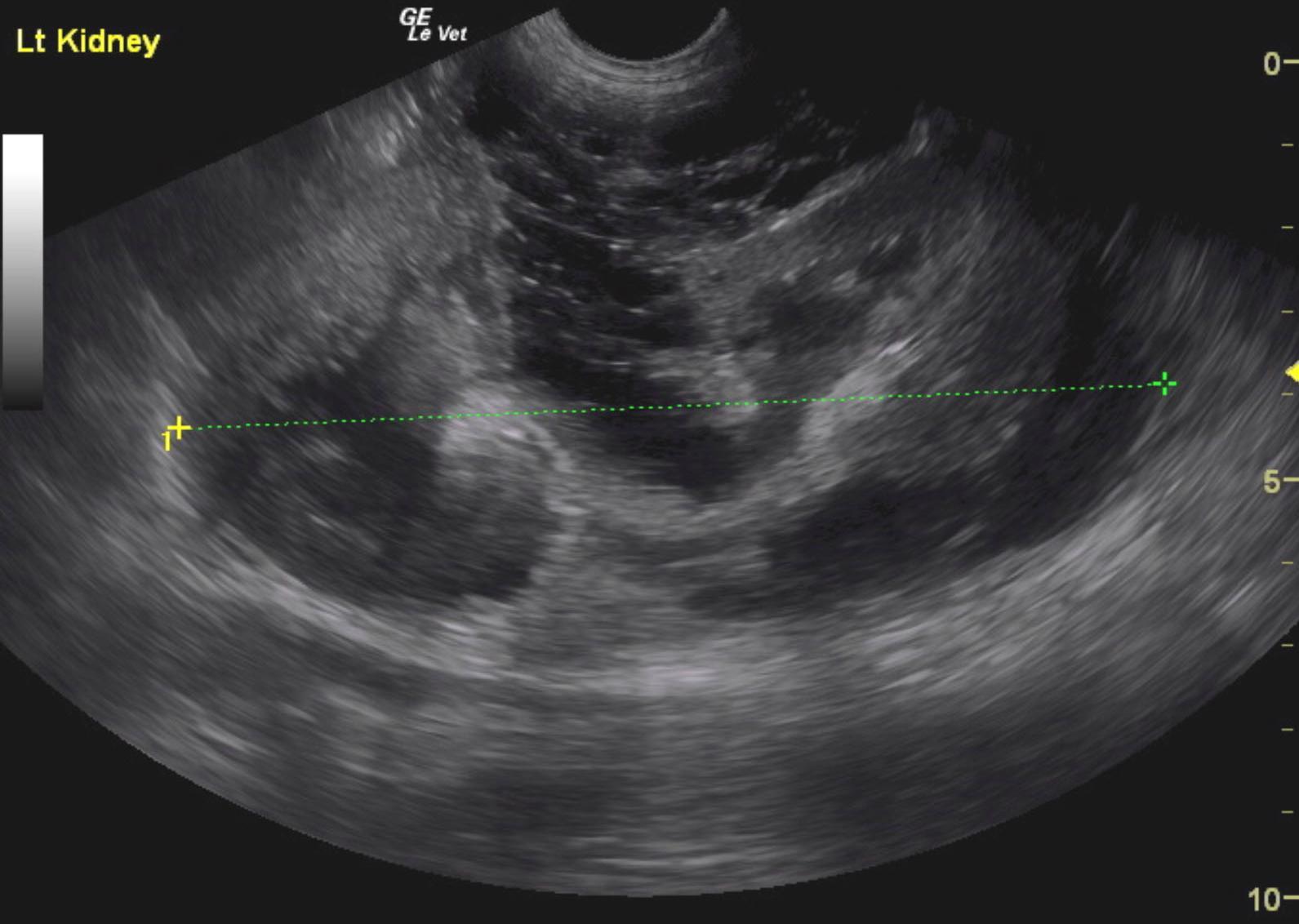
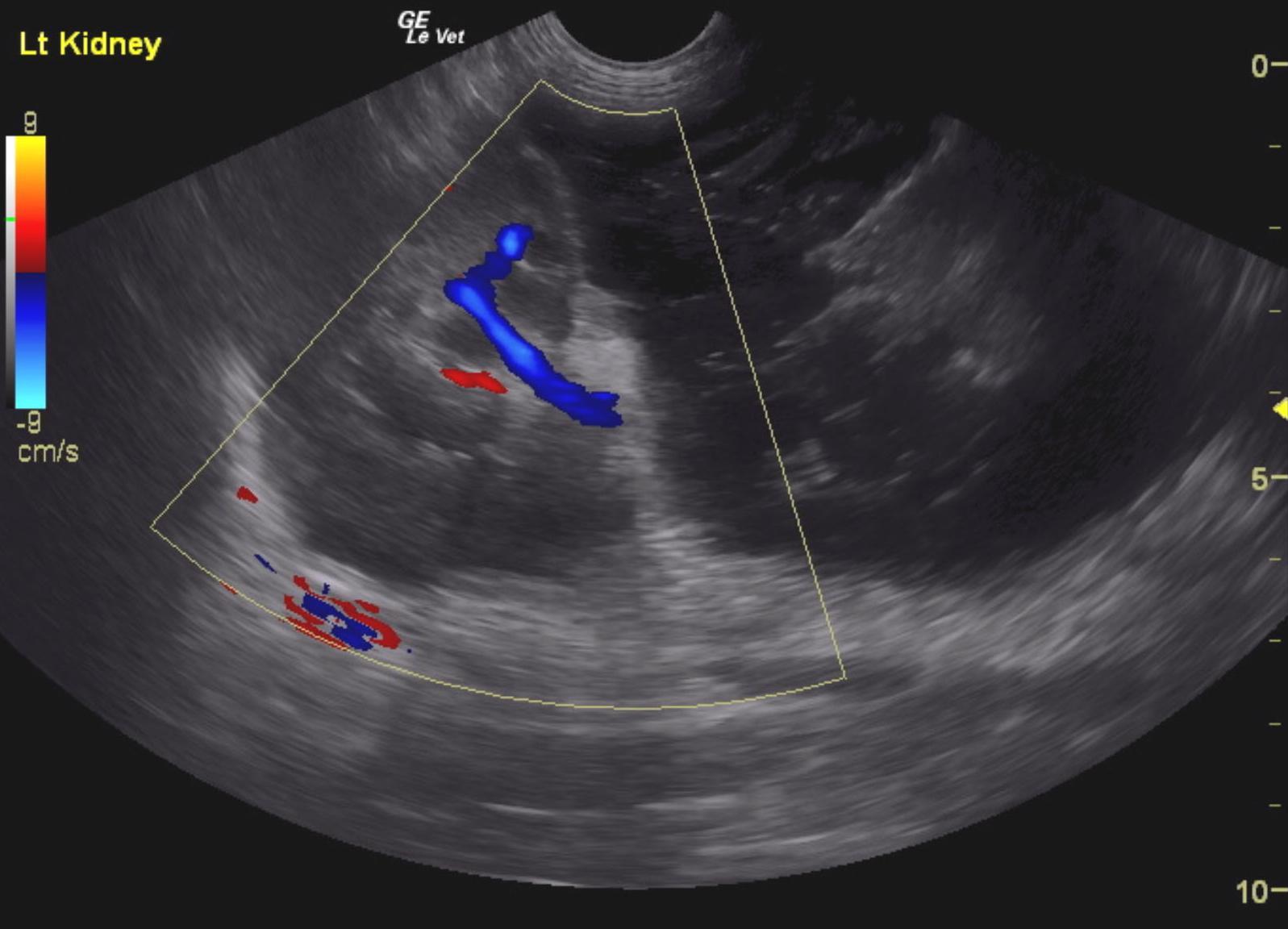
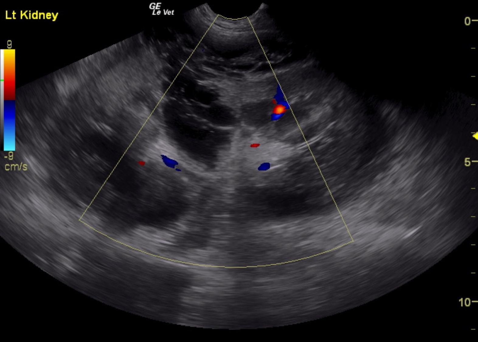
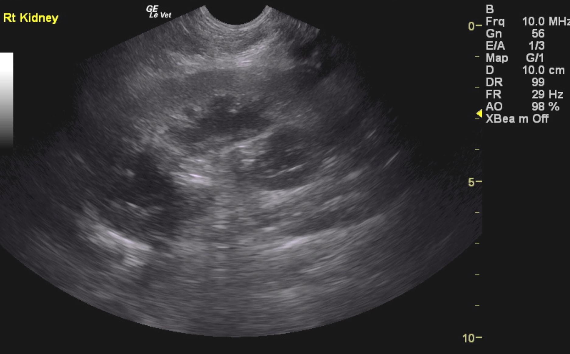
Sonographic Differential Diagnosis: Renal hemorrhage with pericapsular and retroperitoneal fluid and tissue proliferation. Potential clot versus neoplasia and secondary hemorrhage. FNA of the hypoechoic tissue is recommended. Guarded prognosis. Recheck sonogram is recommended in 48-72 hours to assess for any progression or resolution. Neoplastic infiltrates and blood clots can look similar on ultrasound. Doppler assessment could not differentiate one or the other hence the necessity for aspirates.
Sonographic Interpretation:. The left kidney in this patient presented a subcapsular blood clot and hemorrhage escape or possible tumor related escape through the dorsal cortex. This appears to extend outside the capsule. Tissue thickening was noted and extended caudally between the vena cava and aorta. This may represent hemangiosarcoma or other neoplastic event with capsular escape. FNA of the hypoechoic tissue and perirenal space and sublumbar space is recommended to assess for a potential neoplastic event and secondary hemorrhage. Otherwise, exploratory surgery is recommended. However, the patterns extending caudally in the sublumbar and retroperitoneal space will not likely allow for clean resection. The left kidney was severely enlarged at 10.6 cm. Infiltrative process is suspected with potential for a blood clot.