Splenic torsion in a dog.
Sonogram by Andi Parkinson RDMA of Intrapet Imaging, Baltimore, MD, USA. Case evaluation & interpretation by “Team SonoPath.”
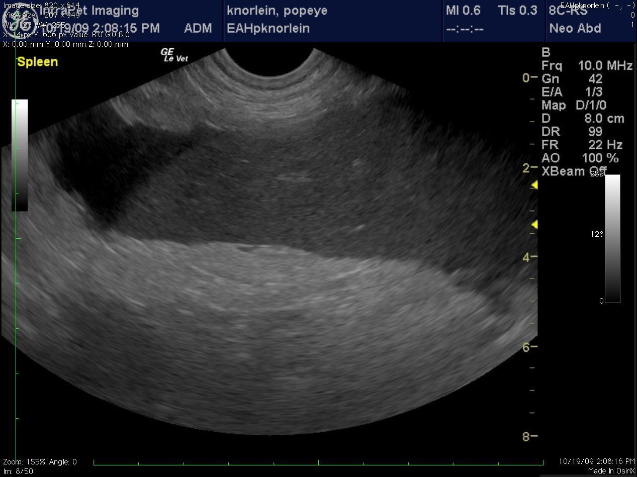
Sonogram (Spleen): “Popeye”
Splenic torsion in a dog.
Sonogram by Andi Parkinson RDMA of Intrapet Imaging, Baltimore, MD, USA. Case evaluation & interpretation by “Team SonoPath.”
Sonogram (Spleen): “Popeye”
History (Lindquist DABVP, Vasquez RVT, Parkinson RDMS): A 6-year-old intact male English Bulldog was presented for intermittent vomiting of 2-3 days’ duration and lethargy. Physical exam found the patient to be tachycardic, with pale mucous membranes, and a palpable mass in the abdomen. In-house serum biochemistry revealed elevated alkaline phosphatase, hypernatremia, and a mild hypochloremia. A decreased RBC, Hct in conjunction with a high MCV was noted, in addition to a neutrophilia, monocytosis and thrombocytopenia. No abnormalities were noted on thoracic radiographs.
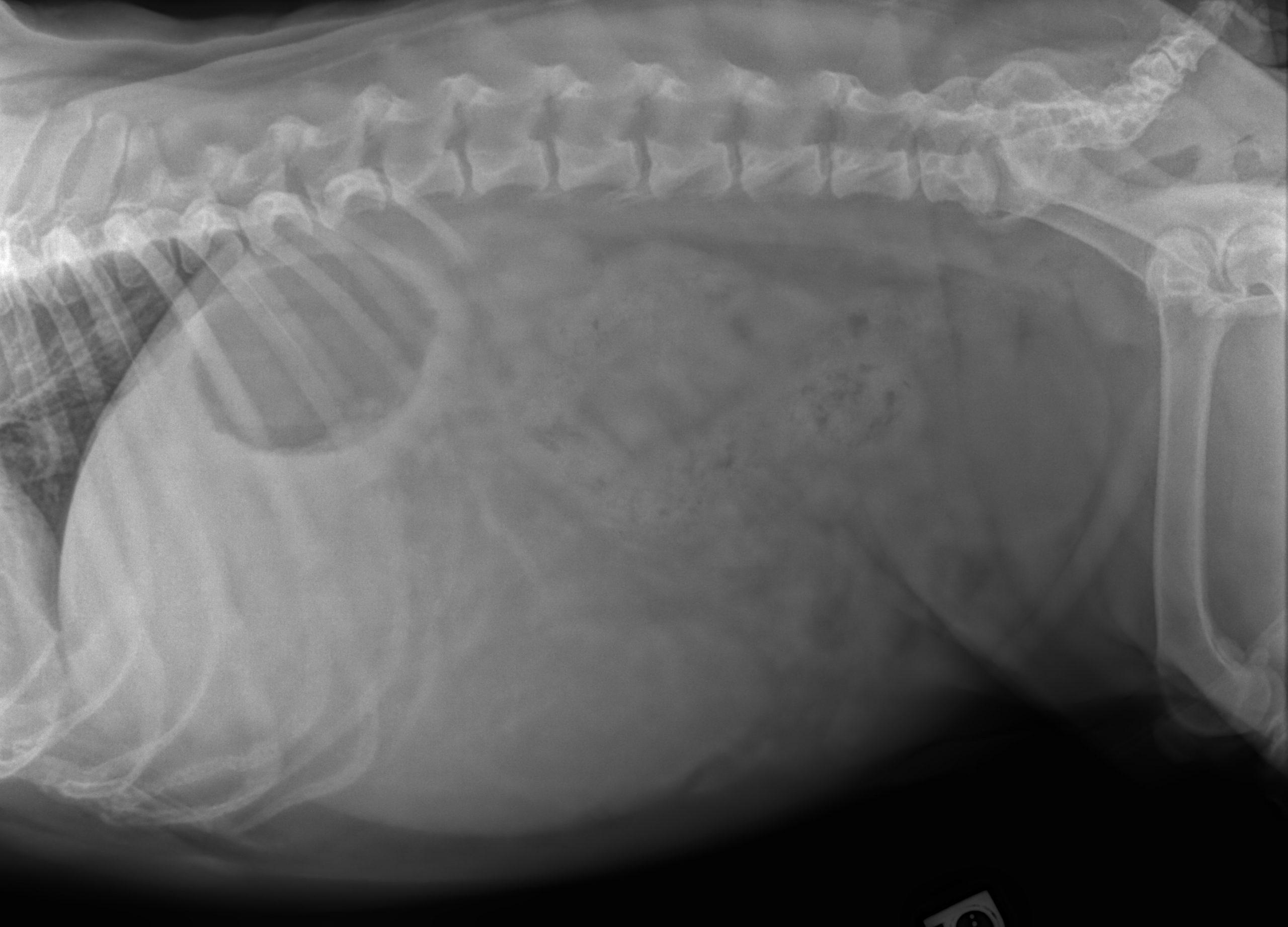
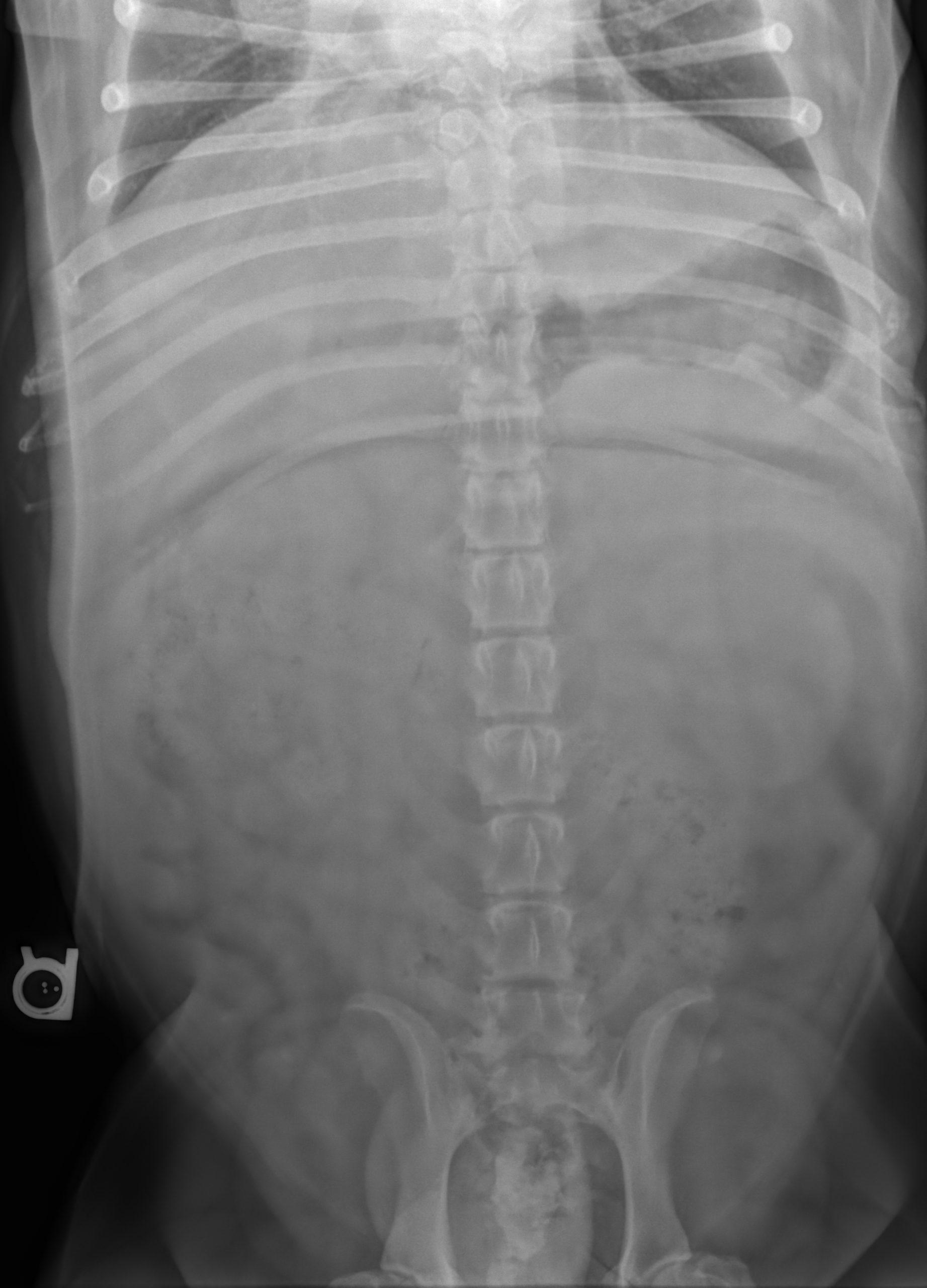
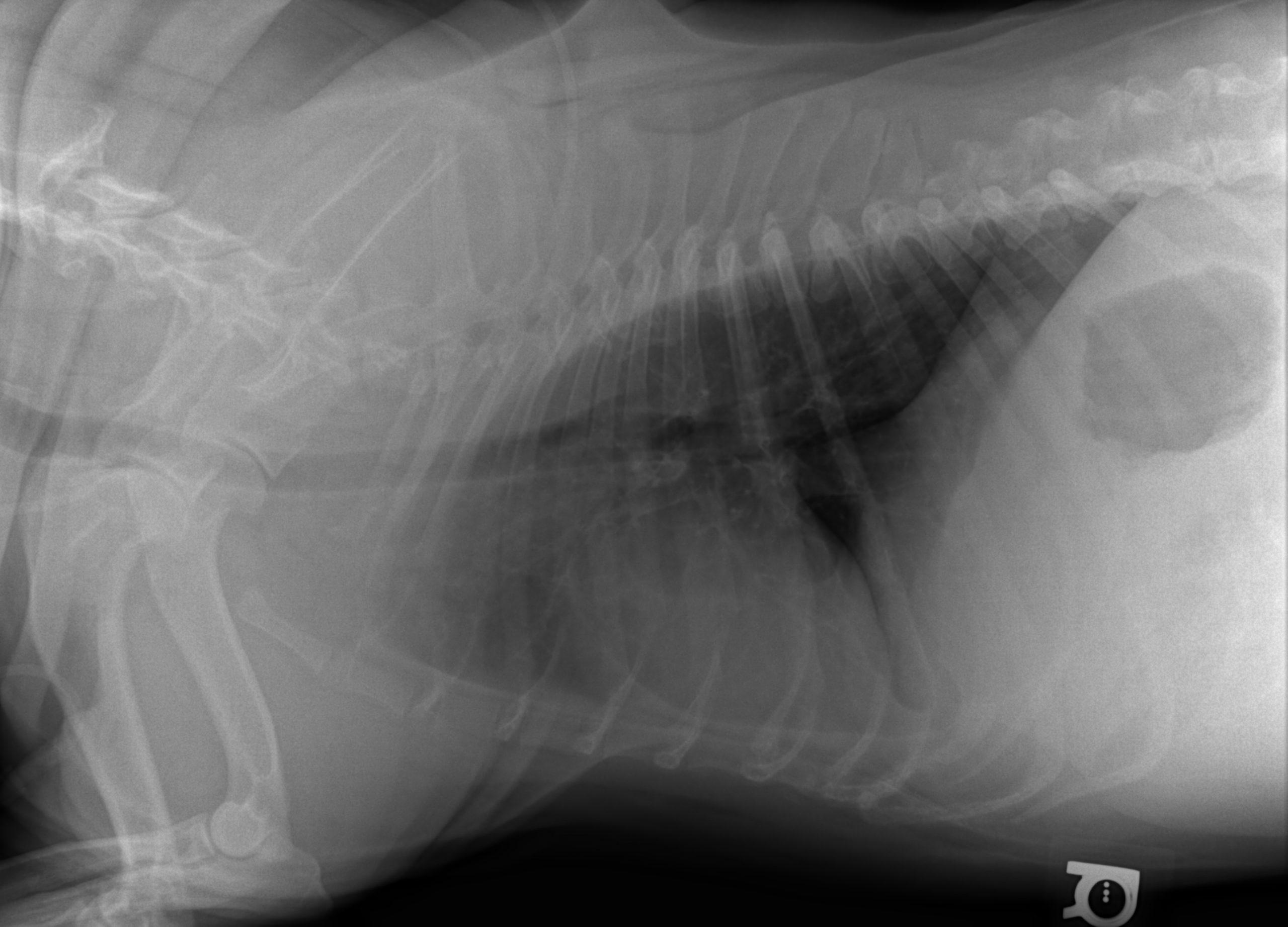
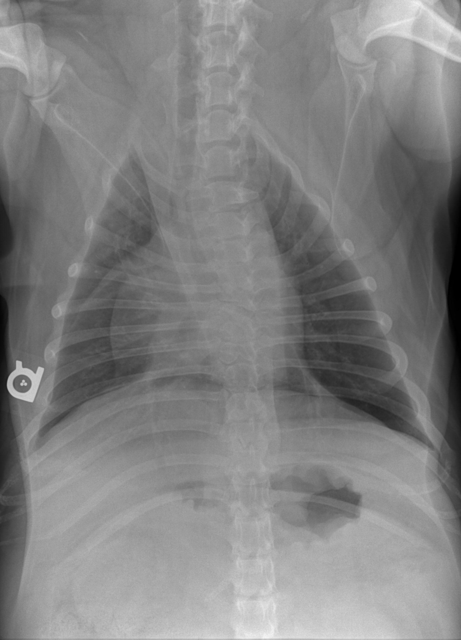
Radiographic Interpretation (Yanik DACVR, Illumipet.com): Abdominal study: Images 1 & 2: Marked splenomegaly strongly suspected to be secondary to torsion. Accompanying infiltrative or neoplastic pathology cannot be ruled out. Small volume of peritoneal effusion and/or inflammation likely secondary to the splenic pathology. Thoracic study: Images 3 & 4: Mild microcardia and underperfused pulmonary vasculature support of hypovolemia. Atypical intrathoracic fat distribution, which is consider incidental. Multiple hemivertebrae.
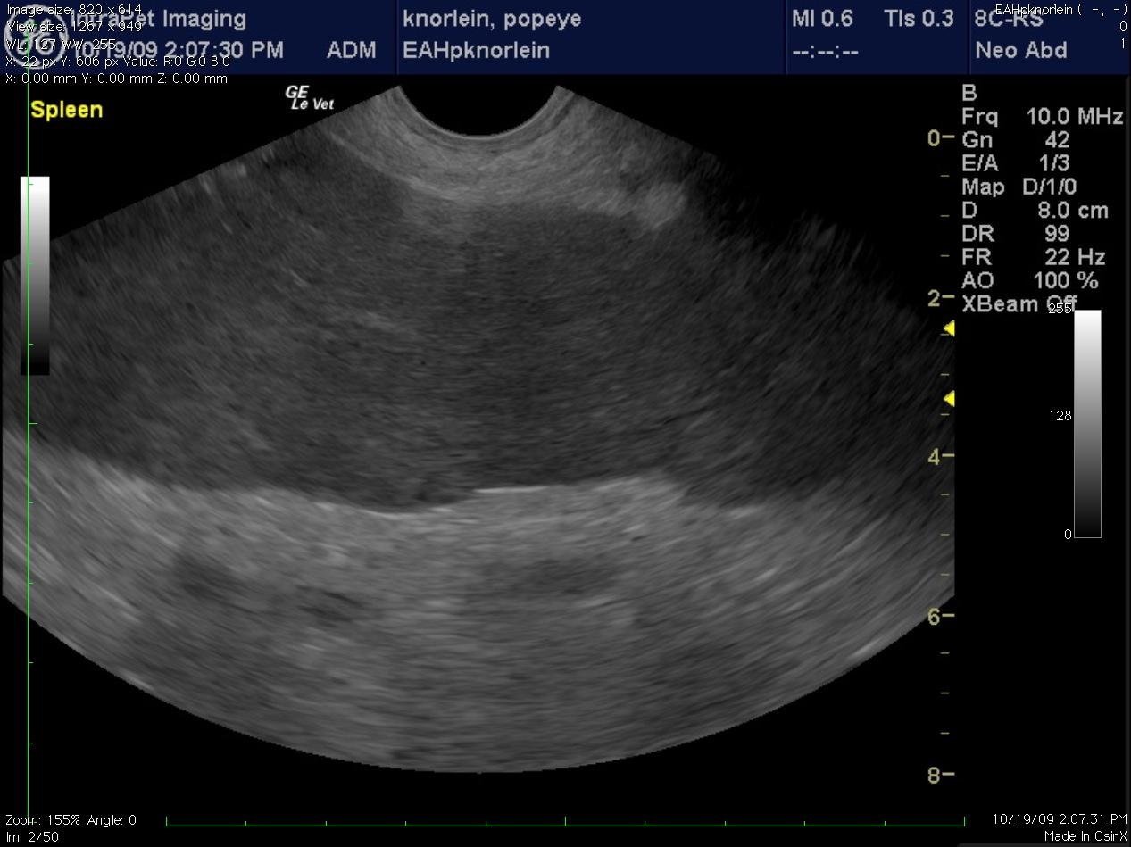
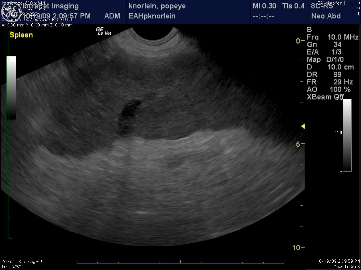
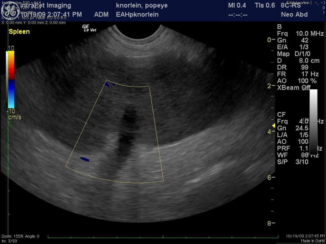
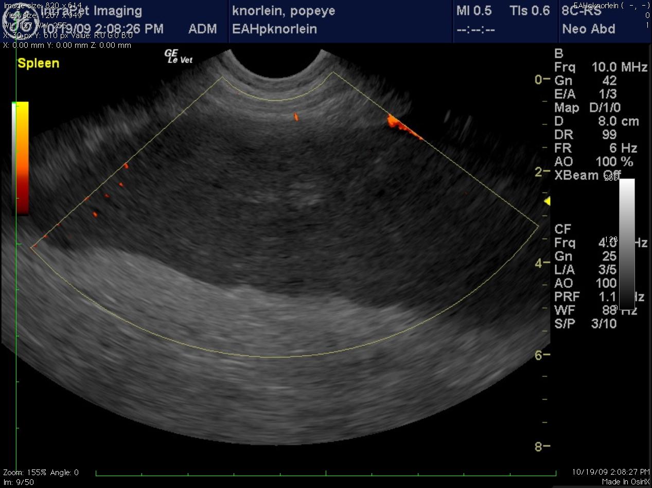
(Yanik DACVR/Lindquist DABVP): Images 5 & 6 demonstrate a canine spleen with mildly heteroechoic, moderately hypoechoic parenchyma, echogenic vascular walls, scalloping capsular contour and increased pericapsular fat echogenicity. Image 7 demonstrates complete lack of blood flow in the hilar vessels given the lack of color flow uptake over the anechoic vessel. Image 8 demonstrates minimal power Doppler uptake over the coarse and micronodular splenic parenchyma. Image 9: The free fluid associated with this presentation indicates either hemorrhage or transudate deriving from an infiltrative process. The video (Image 10) demonstrates a dilated splenic vessel with echogenic content in the far field likely indicating thrombosis given the complete lack of power flow uptake neither within the vessel nor the parenchyma.