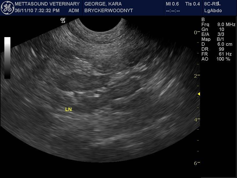
Spontaneous Bowel Torsion On Sonogram. Sonogram performed by Robyn Roberts RDMS of Mettasound Imaging, Austin, Texas, USA. (http://www.mettasound.com/)
Case managed at Bryckerwood Veterinary Hospital, Austin, Texas, USA.
Sonogram: (Abdomen): Kira
Spontaneous Bowel Torsion On Sonogram. Sonogram performed by Robyn Roberts RDMS of Mettasound Imaging, Austin, Texas, USA. (http://www.mettasound.com/)
Case managed at Bryckerwood Veterinary Hospital, Austin, Texas, USA.
Sonogram: (Abdomen): Kira
History: A 3-year-old FS Yellow Labrador had been hit by a car one week prior. The patient sustained pulmonary contusions and was stabilized and discharged. Four days after discharge the patient started intermittent vomiting, diarrhea and inappetance. The patient possibly ate chicken bones around this time frame. The patient was given cerenia for the vomiting. The patient became listless, demonstrated further anorexia and tarry diarrhea. Physical exam revealed a very painful abdomen, capillary refill time of 2 seconds. Bloodwork revealed moderate elevation in ALT, mild decrease in albumin, and no other abnormalities.
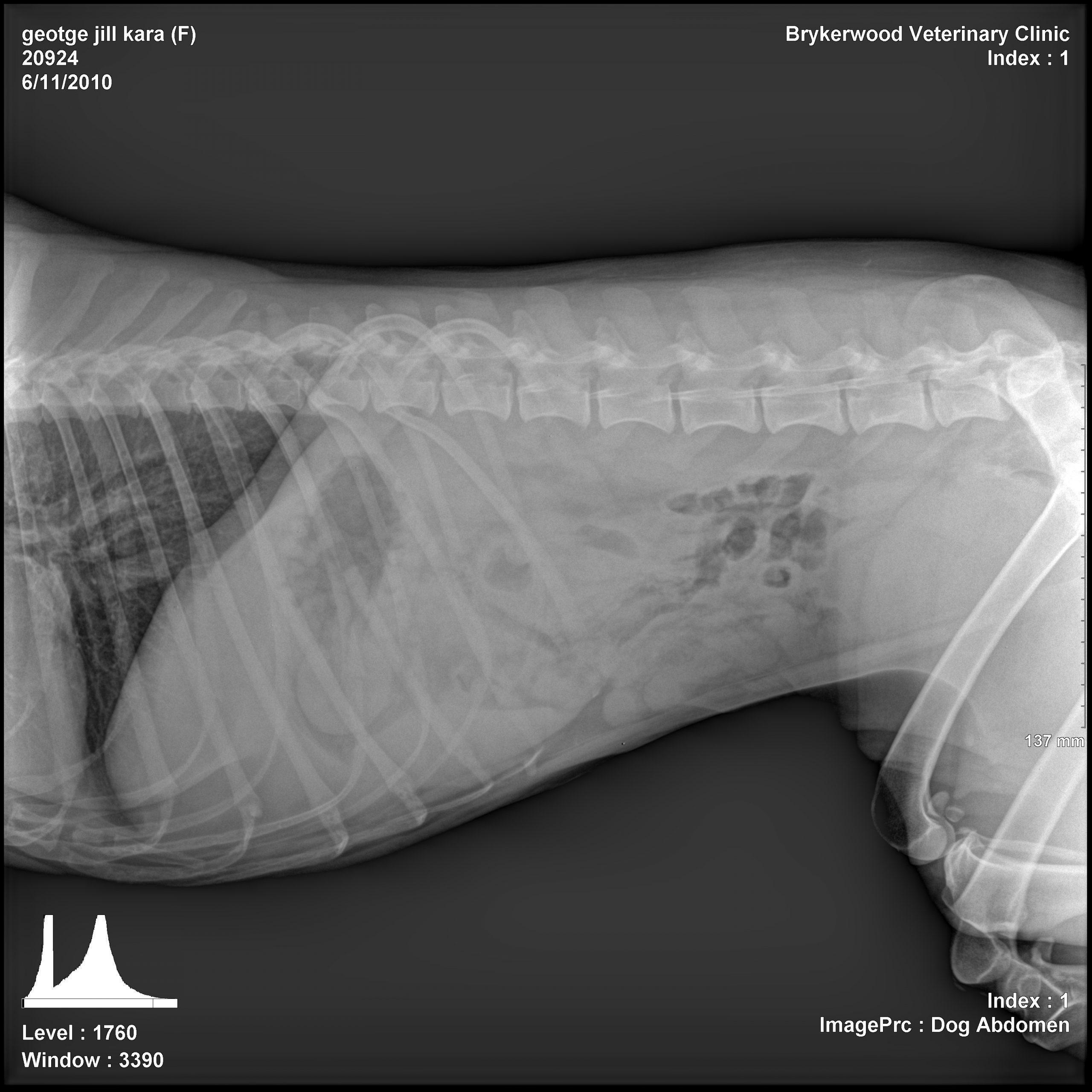
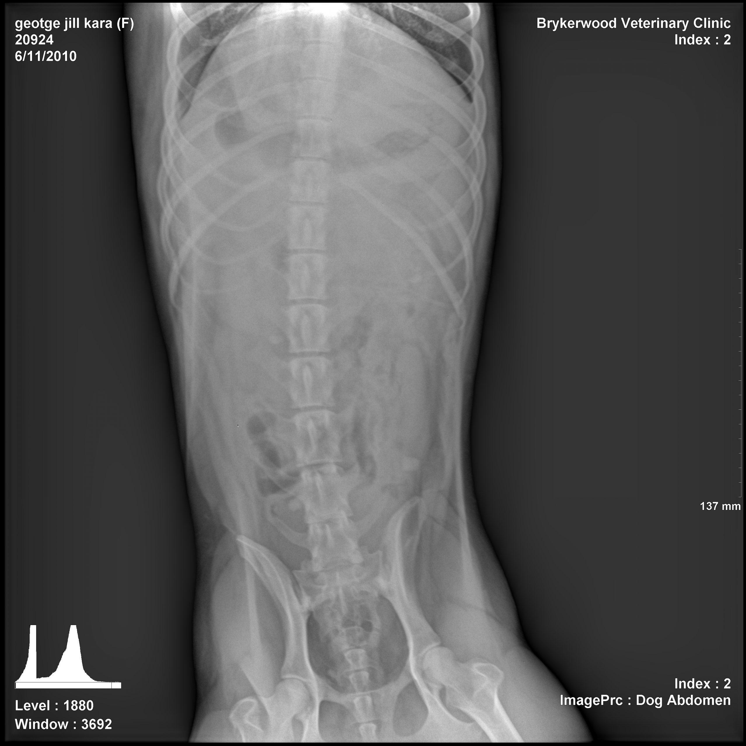
Images 1, 2:
Abdomen:
Thorax: no evidence of the historical pulmonary contusion or other evidence of traumatic thoracic lesions.
Stomach: mild gas and fluid-filled. No foreign body noted.
Small intestines: Mostly clumped caudally in the mid-caudal abdomen. Luminal dimensions of this subset are within normal limits. A second population is suspected in the mid-cranial abdomen on the lateral and ventral-dorsal projections. This subset of intestines have ill-defined serosal margins and measurements are not possible. On the lateral projection one of the loops has a crescent-shaped luminal gas bubble.
The large intestines are uniformly empty, except the terminal descending colon which has faintly mineral-opaque contents. The exact location of the cecum and the relationship o with the aforementioned small intestinal loops can not be determined.
No evidence of free peritoneal air or significant free fluid noted.
Diagnostic Interpretation:
Possible segmental small intestinal dilation; mechanical obstruction (causes include lucent foreign body, intussusception, necrosis, neoplasia)
No distinct gastrointestinal foreign object noted.
No evidence of traumatic thoracic changes or aspiration pneumonia
Testing Considerations:
Abdominal ultrasound and complete thoracic radiographs
Alternatively, pneumocolonography or upper GI series.