This month SonoPath presents an artistic example of sonographic pathology from one of the original consummate GP sonographers & surgeons of our time, Dr. Robert Hylands of Westbridge Veterinary Hospital in Mississauga, Ontario, Canada. Since the 1980s when clinical sonography entered the veterinary scene in serious fashion, Bob has been at the forefront as a luminary and influencer and has always been one of the top clinincal sonographers. Here is just one more rock star example case of bile duct obstruction in a cat with some hidden pathology to accompany it in true Hylands Picasso fashion. Take a look at this sonographic and surgical display and you will see what we mean. 🙂
An 8-year-old DSH feline was presented with an acute presentation of jaundice and decreased appetite. He was also pyrexic (40.6 celsius). Blood chemistry: ALT 669, AST 177, TBil 34, ALKP 48, PLI 50, SDMA 14, T4 22. CBC: normocytic normochromic anemia HCT 28% with reticulocyte 11 (<50) non-regenerative. Urinalysis by cysto; spec. gr 1.047, ph 6.5, Bil3+, prot 2+, RBC>50, WBC 3-5, sq ep 1-5, trans ep 1-5. FELV/FIV NEG.
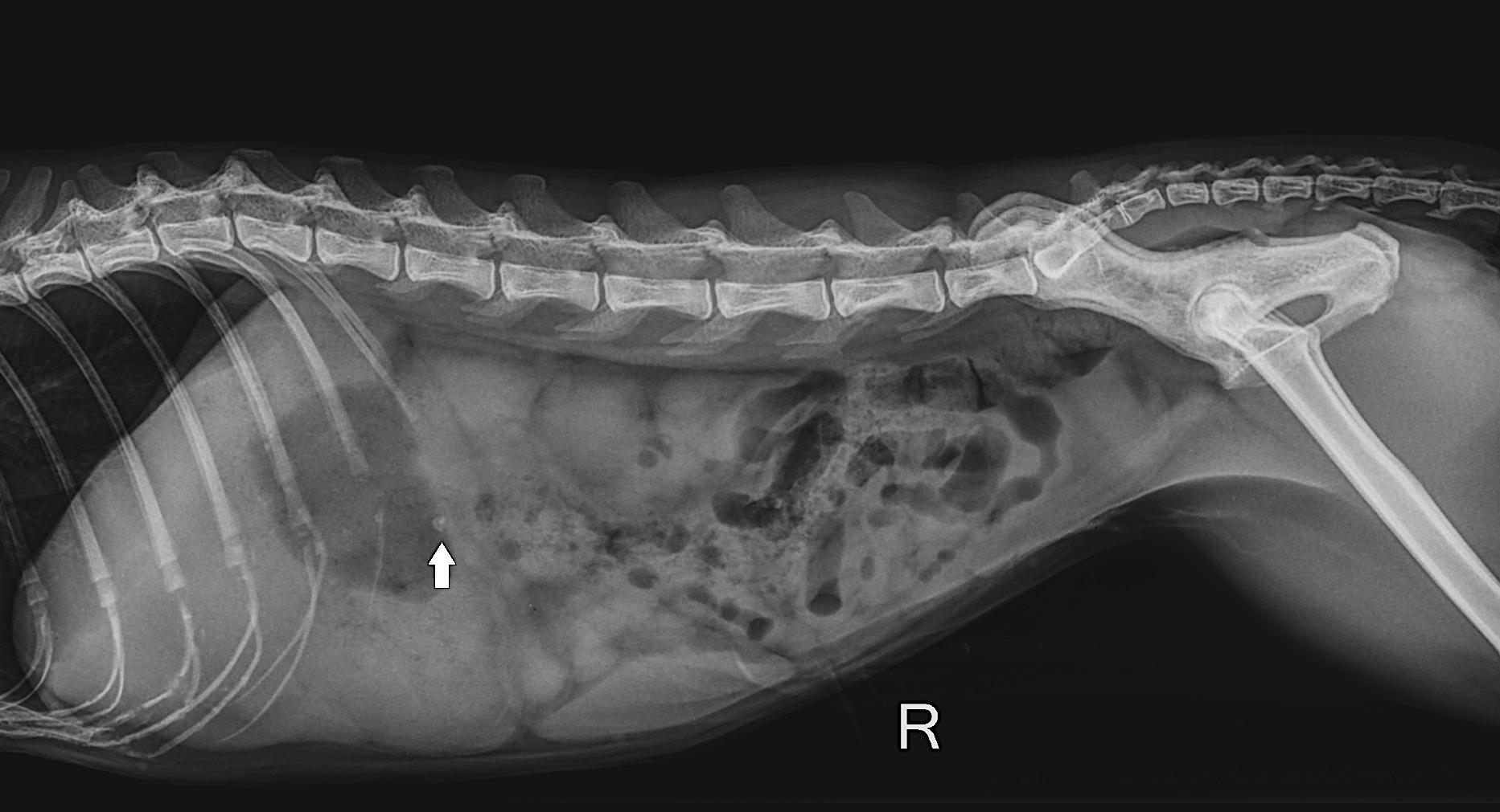
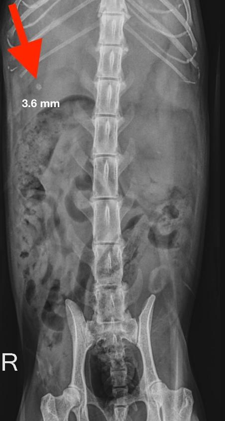
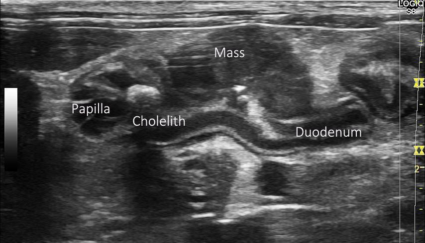
An 8-year-old DSH feline was presented with an acute presentation of jaundice and decreased appetite. He was also pyrexic (40.6 celsius). Blood chemistry: ALT 669, AST 177, TBil 34, ALKP 48, PLI 50, SDMA 14, T4 22. CBC: normocytic normochromic anemia HCT 28% with reticulocyte 11 (<50) non-regenerative. Urinalysis by cysto; spec. gr 1.047, ph 6.5, Bil3+, prot 2+, RBC>50, WBC 3-5, sq ep 1-5, trans ep 1-5. FELV/FIV NEG. Abdominal radiographs clearly demonstrated the presence of a radiopaque density (3 mm) that appears to image in the vicinity of where the major duodenal papilla would be located. The cat has lost almost 0.9 KG (1.9 lbs) over the last year. An abdominal ultrasound was ordered to evaluate the cause of the elevated Tbil and jaundiced appearance of the individual.
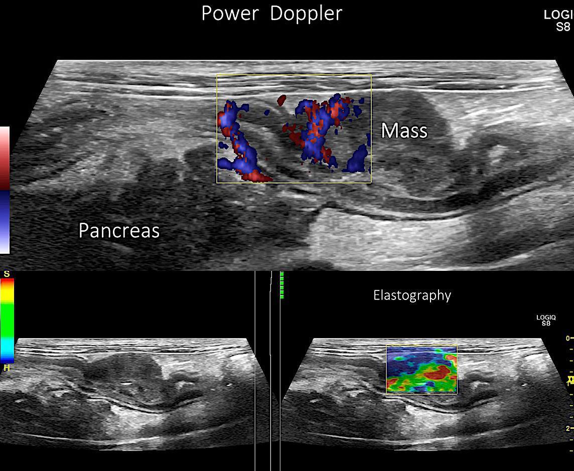
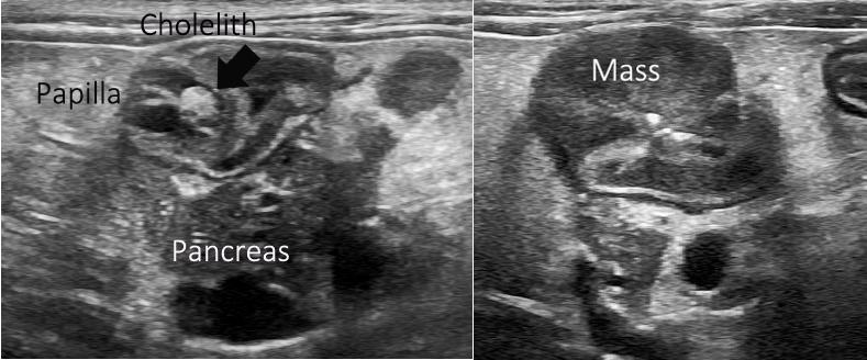
Cholangitis, obstructive cholelith in the papilla, regional peritonitis, pancreatitis, B cell intestinal lymphoma.
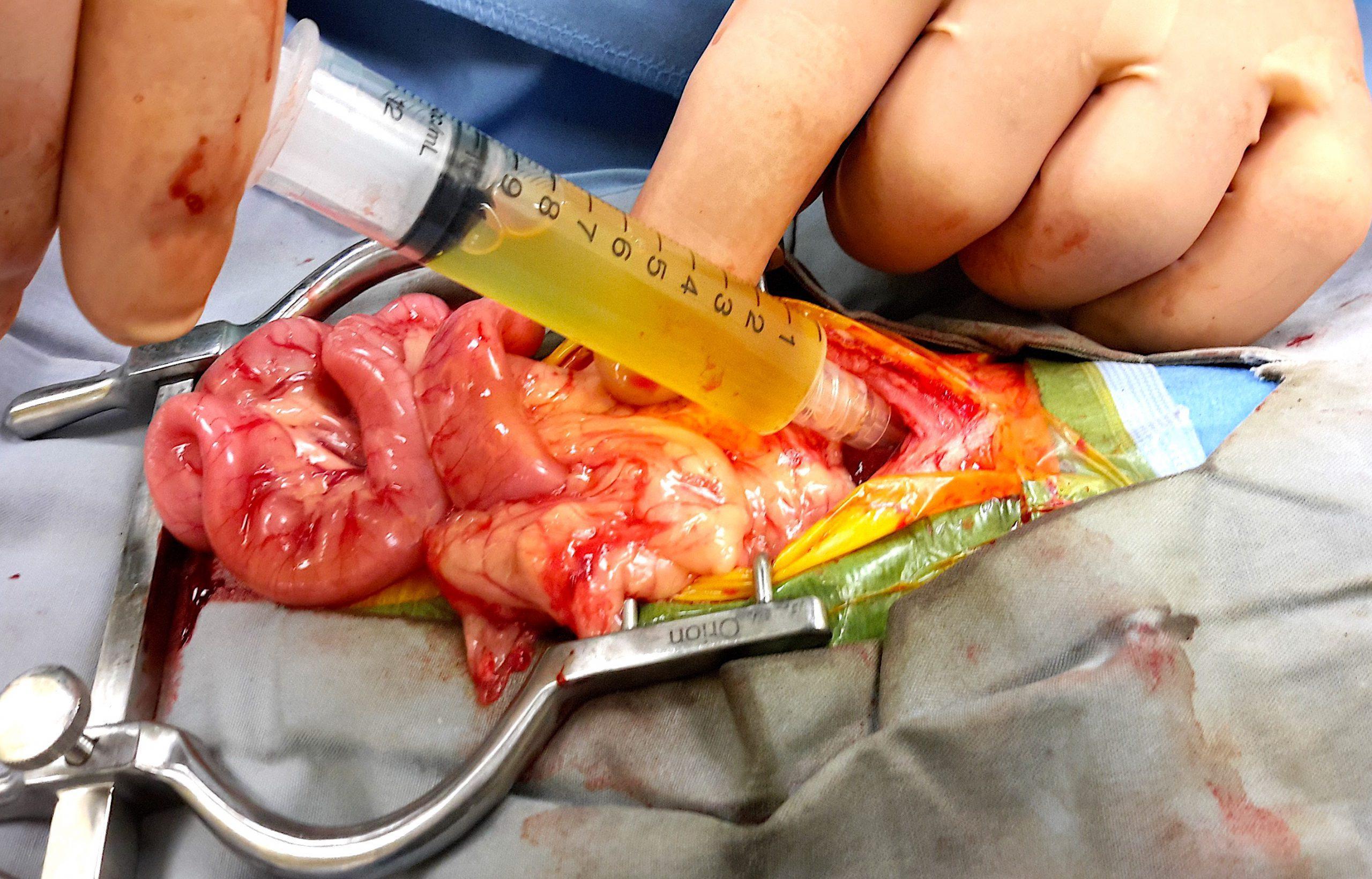
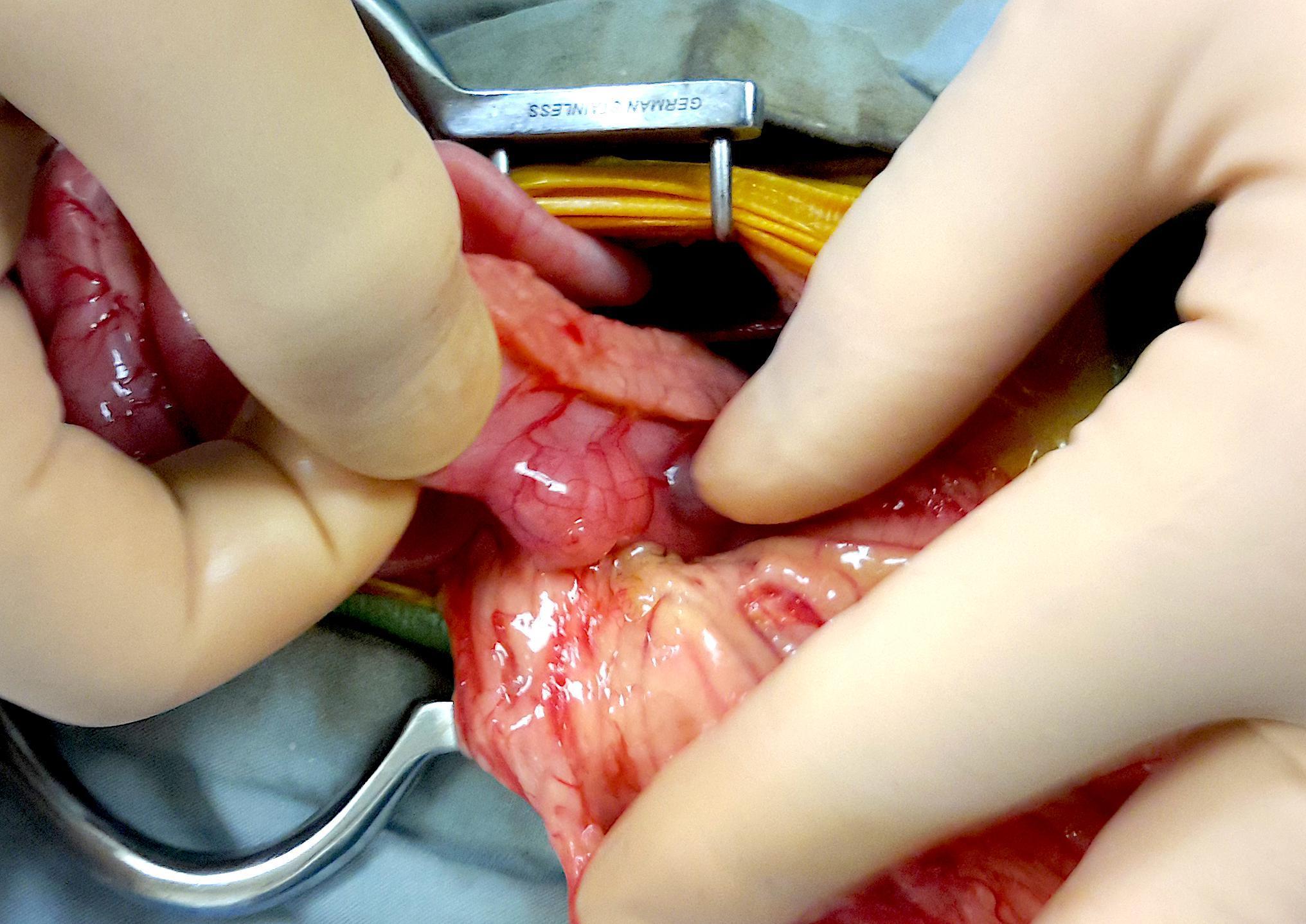
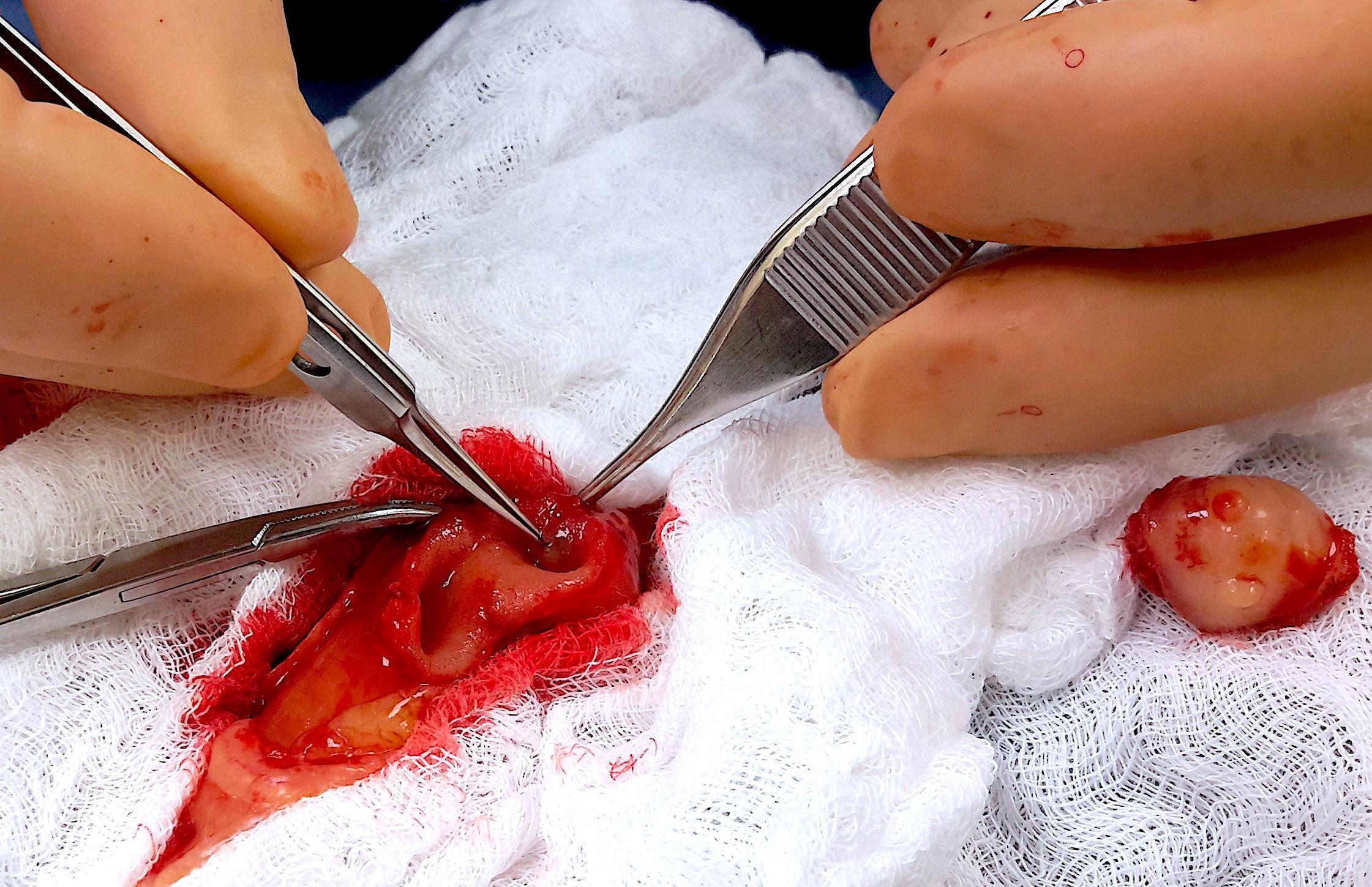
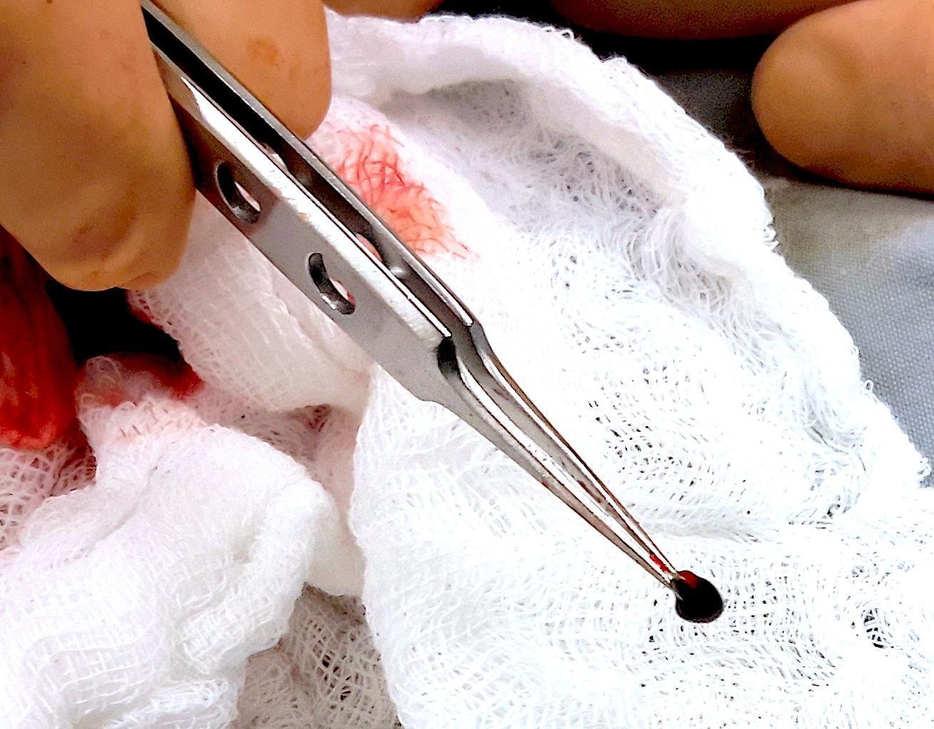
The patient underwent abdominal surgery to remove the cholelith from the papilla. The mass was removed at the same time and a partial intestinal anastomosis performed. This was rather difficult as it was located only millimeters distal to the area of the papilla which needed to be preserved. Biopsies were taken of the kidneys, pancreas, liver and the intestinal mass. Over 15 mls of fluid was aspirated from the CBD to help relieve the intra hepatic accumulations of bile. The cat made a full recovery and was eating well within two days of his surgery. Final biopsy results were the following: neutrophilic cholangiohepatitis, interstitial nephritis, fibrosing pancreatitis, large b cell intestinal lymphoma. The owners opted for no further treatments for their cat as they were worried that the medications may affect their pregnancy. The cat succumbed 5 months later at an emergency clinic from unrelated respiratory issues.