Patient presents for suspicion of possible Cushing’s. LDDST did not support a diagnosis of Cushing’s, however the chemistry panel does have generalized increased liver values; all considered mild-mod. ALT, AST, ALKP all mildly increased. Mild thrombocytosis – likely excitement. SDMA mildly increased. BUN/Crea normal. Abdominal ultrasound was recommended.
Patient presents for suspicion of possible Cushing’s. LDDST did not support a diagnosis of Cushing’s, however the chemistry panel does have generalized increased liver values; all considered mild-mod. ALT, AST, ALKP all mildly increased. Mild thrombocytosis – likely excitement. SDMA mildly increased. BUN/Crea normal. Abdominal ultrasound was recommended.
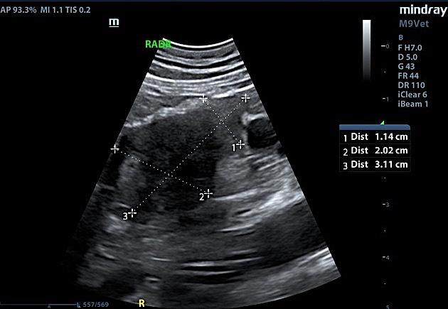
Right adrenal mass with possible early phrenic +/- caval invasion. No obvious large invasion, appears potentially resectable.
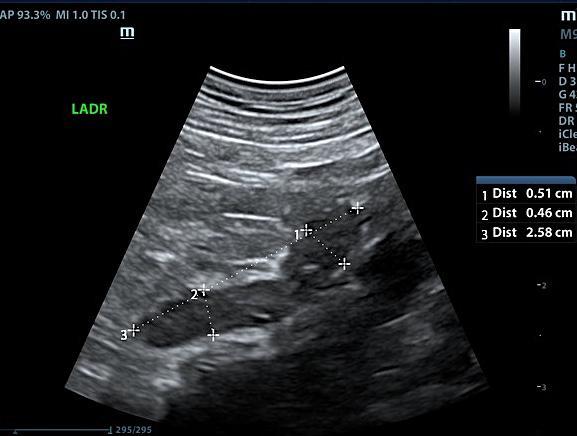
The left adrenal gland was visualized and recognized as having normal shape, size, position and echogenicity for this breed. The phrenic vasculature, glandular echogenicity and detail were unremarkable. Capsule, cortex, and medullary definition were normal for this age patient. The left adrenal gland measured 2.58 x 0.46 cm at the caudal pole and 0.51 cm at the cranial pole. The right adrenal gland had a disrupted curvilinear pattern with microcystic changes. The right adrenal gland measured 3.1 x 2.02 cm at the cranial pole and 1.14 cm at the caudal pole. Capsular expansion was noted. The right adrenal gland was moderately vascular. The right adrenal gland impinges upon the vena cava without obvious invasion. Minor invasion into the phrenic cannot be ruled out. The right adrenal gland appears potentially resectable. The vena cava prior to and after the adrenal gland did not appear invaded.