A 4-year-old intact male labrador retriever was presented for depression, weight loss, intermittent vomiting bile, decreased appetite, and greenish, mucoid ocular discharge. Blood work revealed mild leukocytosis and mild hyperglobulinemia.
A 4-year-old intact male labrador retriever was presented for depression, weight loss, intermittent vomiting bile, decreased appetite, and greenish, mucoid ocular discharge. Blood work revealed mild leukocytosis and mild hyperglobulinemia.
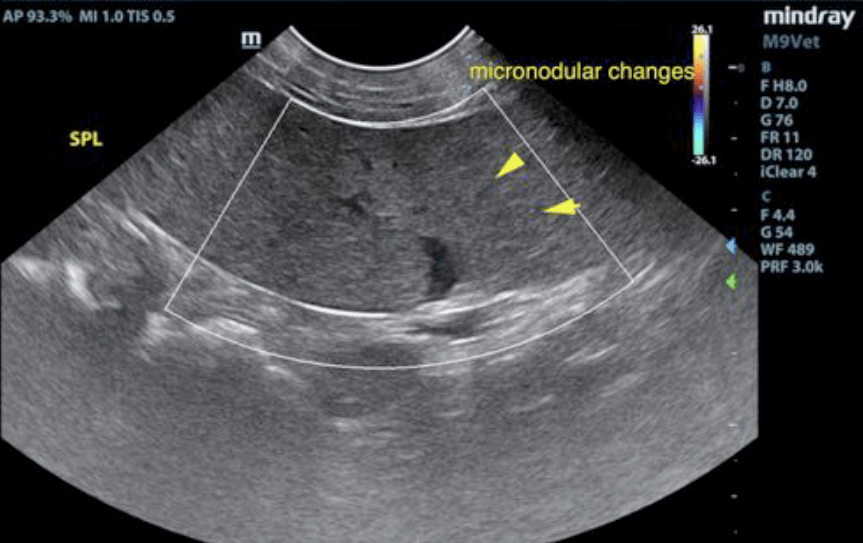
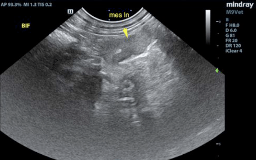
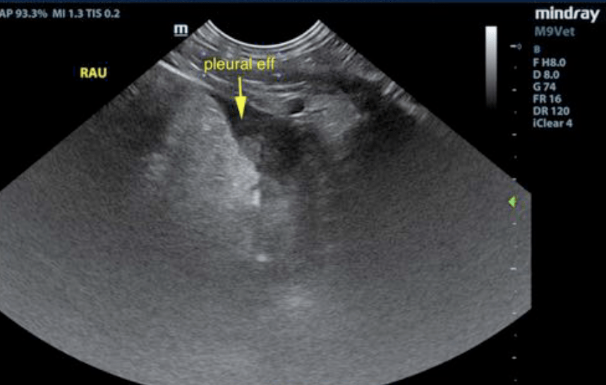
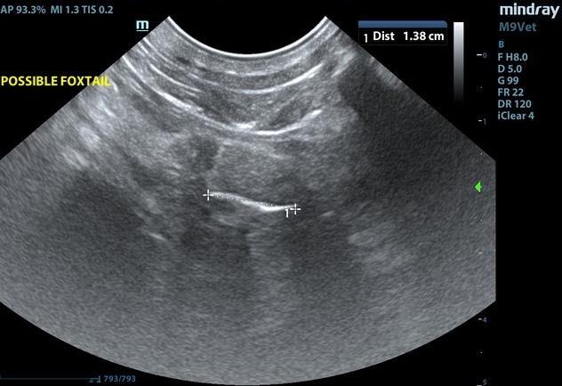
BPH prostate. Enlarged, scalloping spleen. Mesenteric lymphadenopathy. Foxtail foreign body suspected in lung. Lung necrosis and pleuritis is suspected.
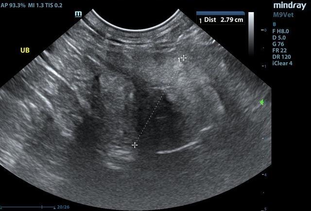
The prostate was uniformly enlarged with lobar swelling appearing to impinge upon the urethra and mildly deviate the descending colon. The spleen was slightly enlarged with minor scalloping contour. The mesenteric lymph nodes were mildly enlarged. The length to width ratio was maintained and measured 2.0 x 1.0 cm. Rapid view of the heart revealed no evidence of pathology. The extracardiac space revealed a 5.0 cm lung consolidation with regional pleural effusion. A 1.5 cm linear structure was noted in the lung field.
CT evaluation of the chest would be ideal. Ultrasound-guided FNA of the pleural effusion and area of the lung is recommended to ensure that this is not a neoplastic presentation. FNA of the spleen would also be warranted to assess for splenitis. There may be multiple issues in this patient including suspected fox tail; however, underlying neoplasia is a potential. Sampling and CT evaluation +/- thoracotomy would be warranted. The patient was transferred to a specialty facility for surgical removal of the foxtail and concurrent lung lobectomy. The patient recovered and is doing well.