Innocent murmur… I think not! Here is a case where acting on a heart murmur, no matter the age or grade, is a prudent choice in clinical practice. A young, rambunctious boxer was presented for an echocardiogram and ECG, for a low grade (1/6) heart murmur that had continued to be auscultated after several puppy visits to her veterinarian. No obvious clinical signs were present, other than panting, but not unusual for an exuberant puppy in a clinic setting! Many thanks to Dr. Shelly Knopsnyder of East Lane Veterinary Hospital and her awesome team in managing this case. SDEP-certified sonographer, Amanda Lacey of Animal Sounds NW provided the diagnostic images for this case study.
A 1.5-year-old, FS, Boxer was presented for an initial puppy wellness visit and a grade 1/6 cardiac murmur was detected; noted PMI (point of maximal impulse) right cranial. A grade 1-2/6 cardiac murmur was detected at several follow-up visits. The patient underwent ovariohysterectomy without event. More recently the patient was presented for further cardiac evaluations. PE found the patient with a heartrate of 140, panting but with no increased respiratory effort, and synchronous pulses. BP: 111/51, 95/53 MAP 67, 119/66 MAP 78. 2 ECG strips were submitted.
A 1.5-year-old, FS, Boxer was presented for an initial puppy wellness visit and a grade 1/6 cardiac murmur was detected; noted PMI (point of maximal impulse) right cranial. A grade 1-2/6 cardiac murmur was detected at several follow-up visits. The patient underwent ovariohysterectomy without event. More recently the patient was presented for further cardiac evaluations. PE found the patient with a heartrate of 140, panting but with no increased respiratory effort, and synchronous pulses. BP: 111/51, 95/53 MAP 67, 119/66 MAP 78. 2 ECG strips were submitted. The first strip (taken under sedation with butorphanol) showed periods of sinus rhythym and periods that appeared to be a high grade second degree AV block (ventricular rate 40-100 bpm). The second strip showed a sinus rhythm (rate 114 bpm) with intermittent single premature ventricular complexes (RBBB morphology) once sedation had worn off and patient was stimulated.
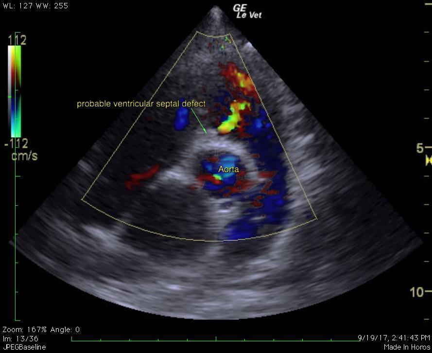
Venticular septal defect.
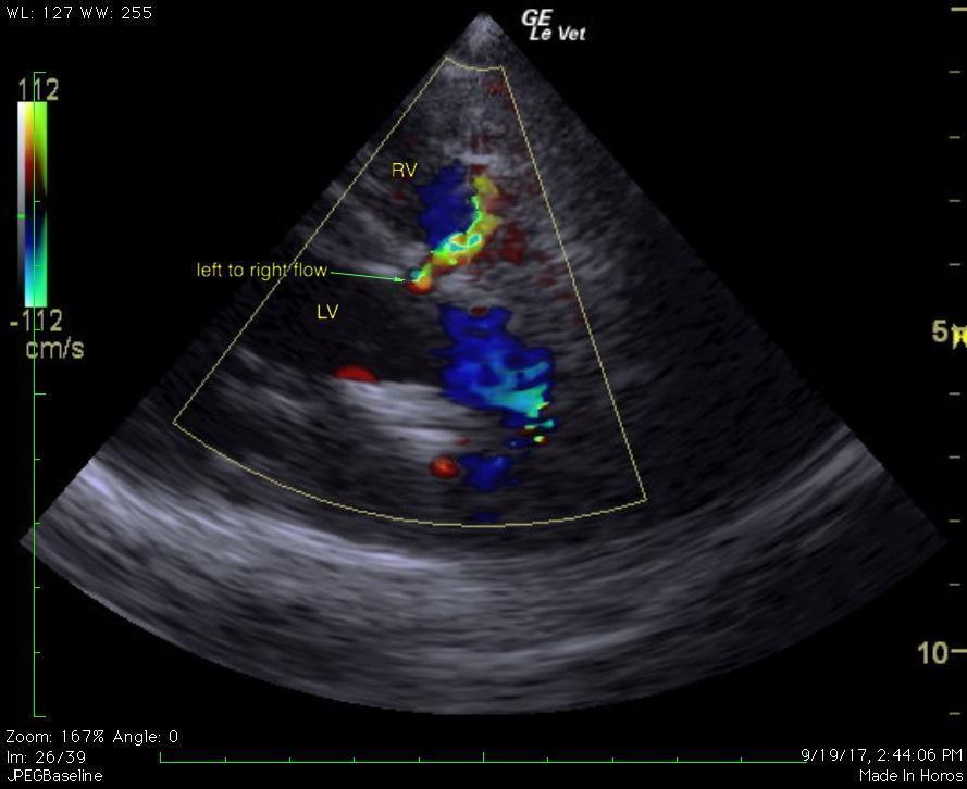
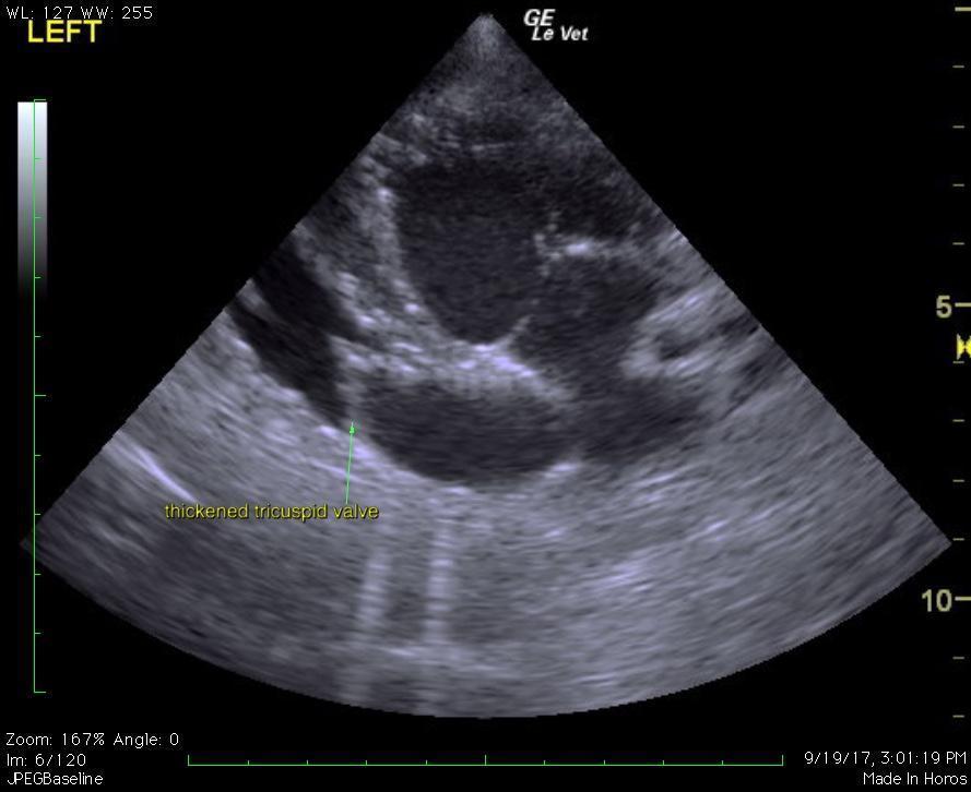
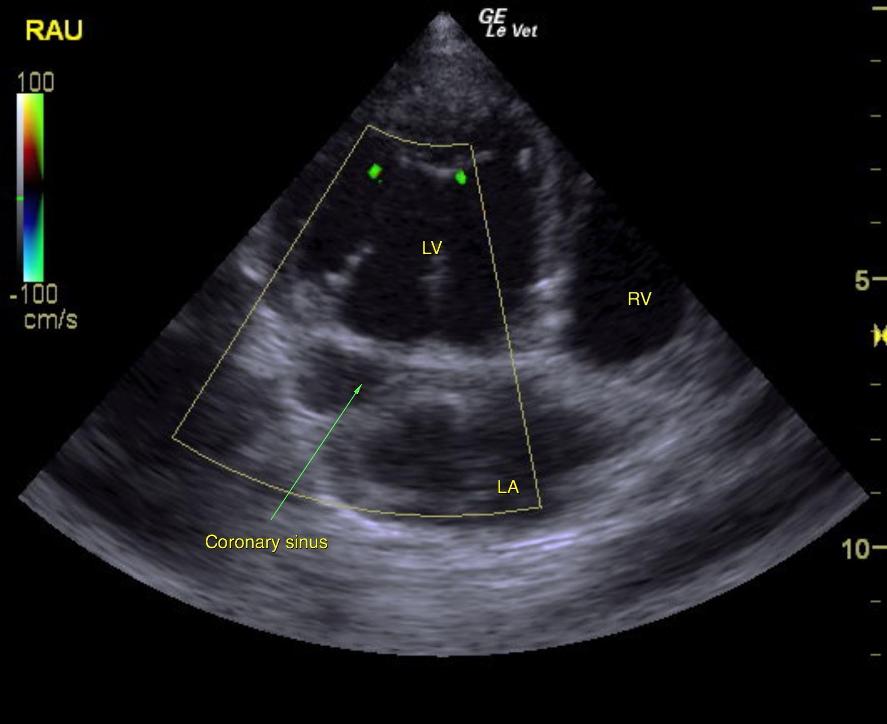
The murmur appears to be secondary to a small left to right shunting ventricular septal defect (~5mm), suspected restrictive physiology. Probable tricuspid valve dysplasia. Dilated coronary sinus (Ddx persistent left cranial vena cava vs. anomalous pulmonary venous return vs. other). Ventricular premature complexes. Possible high grade AV block during sedation (Ddx high vagal tone vs. myocarditis vs. idiopathic nodal fibrosis vs. other).
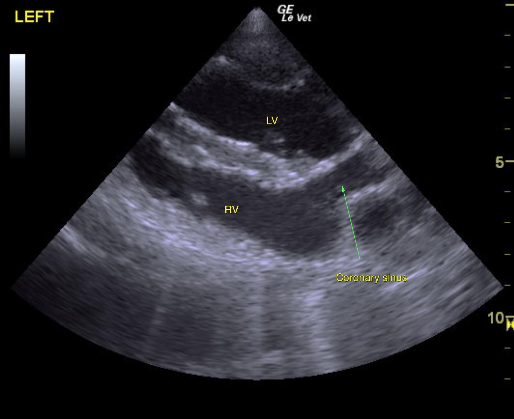
A sinus rhythm was observed during the echocardiogram. There is a dilated coronary sinus. Trivial mitral regurgitation is suspected. The tricuspid valve leaflets are thickened and the septal leaflet is thethered in some views, although there is no tricuspid regurgitation. There is an area of left to right flow (beneath the junction of the right and non-coronary aortic cusps high in the ventricular septum).
Placement of a 24-hour Holter monitor was advised to better assess the arrhythmia documented in the ECG, which is unlikely to be related to the VSD. If concurrent AV block and ventricular arrhythmias are diagnosed, a myocarditis may be present; in this case tick titers, protozoal titers, and potential for treatment with doxycycline would be indicated. The dilated coronary sinus may be related to a persistent left cranial vena cava or anomalous pulmonary venous drainage. A bubble study in the left cephalic vein would confirm the suspicion of a persistent left cranial vena cava. This is typically a benign congenital change. The lack of tricuspid regurgitation is a favorable finding. No cardiac medications appear indicated at this point, given the lack of clinical signs and lack of congestive heart failure on radiographs. With a small VSD the prognosis is typically good; recheck echocardiogram is 6-9 months was recommended. Due to the patient being clinically normal at home. as she was prior to the ultrasound, owners elected to not pursue further care except for recheck/echo/ECG that was recommended by the cardiologist.