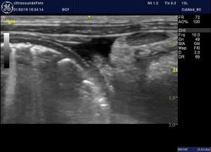

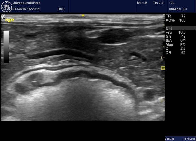
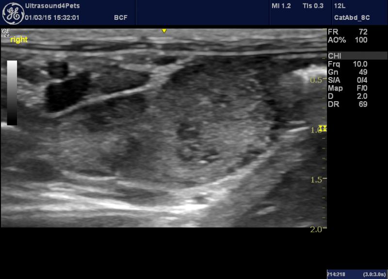
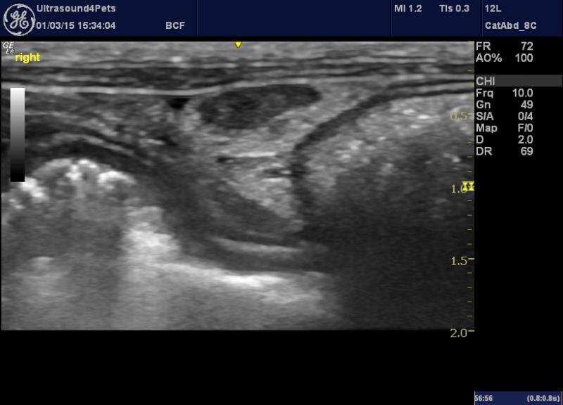
images above 1) effusion 2) L pancreas 3) jejunal LN 4) colic LN
This is an adult oriental cat with a long history of ill-thrift, diarrhoea and occasional vomiting.
I don’t think I’ve seen mesenetric LNs which look quite like this before. They are all peppered with hypoechoic foci -each of which contains a hyperechoic ‘fleck’.
There are diffuse changes in the jejunal mucosa (increased echogenicity, no loss of layering). Small effusion. Remainder of abdo examination unremarkable.
Would anybody read anything specific into this LN appearance? I’m presuming that they are probably foci of necrosis or microabscesses???
Hoping to get some histo and find out for sure in due course.
Thanks
Roger
6 responses to “unusual jejunal and colic lymphadenopathy in a cat”
Microabscesses, granulomatous
Microabscesses, granulomatous disease, lsa, mct ca all do this. Needs a needle. The free fluid is likely from lymphatic strangulation and modified transudate. If you get any fluid or fna with a touch of saline in the syringe you can culture them. Beautiful images roger
Microabscesses, granulomatous
Microabscesses, granulomatous disease, lsa, mct ca all do this. Needs a needle. The free fluid is likely from lymphatic strangulation and modified transudate. If you get any fluid or fna with a touch of saline in the syringe you can culture them. Beautiful images roger
Thanks Eric
Thanks Eric
Thanks Eric
Thanks Eric
Would also recommend fluid
Would also recommend fluid cytology as it may not be secondary to lymphatic strangulation.
Would also recommend fluid
Would also recommend fluid cytology as it may not be secondary to lymphatic strangulation.